
Figure 7.
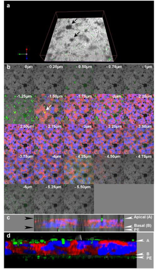
Distribution of 300nm long p(4VP)-MWCNTs across a TT1 cell monolayer at 24h following apical application. (a) Confocal image showing the p(4VP)-MWCNTs (green) at the basal side of the cell, within the supporting polyester membrane (PE), many located at membrane pores (section −4.75 to −5μm in panel b). The 4VP-MWNTs are green in the reflecting channel and they can be seen as black patches in the transmission channel. (b) Translocation of p(4VP)-MWCNTs across the TT1-monolayer (blue=nuclei and red=plasma membrane); the stack was 0.25 μm thick and the value on each image indicates focal height of the image along the z-axis. Aggregated p(4VP)-MWCNTs (green and black patches in b) were observed within the TT1-monolayer and some MWCNTs co-localised with the cell cytoplasm (yellow in panel b at z= −1.5 and −1.75). (c-d) 3D images of stack images from 4 channels: green, red, blue with (c) /without transmission channel (d) were combined and illustrated in yz-plane. The green layer on the top and at the basal side of cell monolayer (black arrows in c) indicate the aggregated p(4VP)-MWCNTs adhered on the apical side of the cell and the p(4VP)-MWCNTs crossed the monolayer which were trapped by the supporting transwell membrane (PE), respectively.