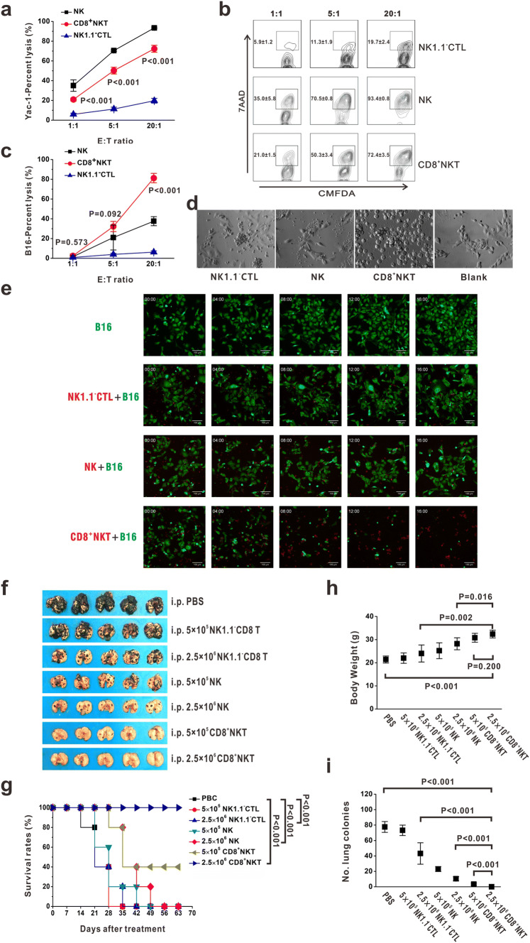
Fig. 1.
CD8+NKT-like cells kill tumor cells similar to NK cells. a, b CMFDA-stained Yac-1 cells were co-cultured with either CD8+NKT-like cells or NK cells or NK1.1−CTLs at E:T ratios of 1:1, 5:1, and 20:1. After 24 h, 7-AAD was added and the apoptotic cells were detected by flow cytometry. Statistical data (a) and the corresponding flow cytometry charts (b) are shown. c, d CMFDA-stained B16 cells were co-cultured with either CD8+NKT-like, NK cells or NK1.1−CTLs at E:T ratios of 1:1, 5:1, and 20:1. After 24 h, 7-AAD was added and the apoptotic cells were examined by flow cytometry (c). Morphology of the co-culture system at the E:T ratio of 5:1 was observed with microscopy (d). e Dynamics of the killing process of B16-GFP cells (green) by CD8+NKT-like cells (red), NK cells (red) or NK1.1−CTLs (red) are shown. f, i. A total of 5 × 10⁴ B16 melanoma cells were intravenously inoculated into C57BL/6 mice. After 12 h, 5 × 10⁵ (low-dose group) or 2.5 × 10⁶ (high-dose group) CD8+NKT-like cells or NK cells or NK1.1−CTLs were used for peritoneal adoptive transfer. Lung metastasis (f), survival rates (g), body weights (h), and number of lung metastatic colonies (i) for each group are shown. In vitro experiments were performed in triplicate and repeated three times with similar results. In vivo experiments were repeated three times. n = 5 for each repeat

