Figure 8.

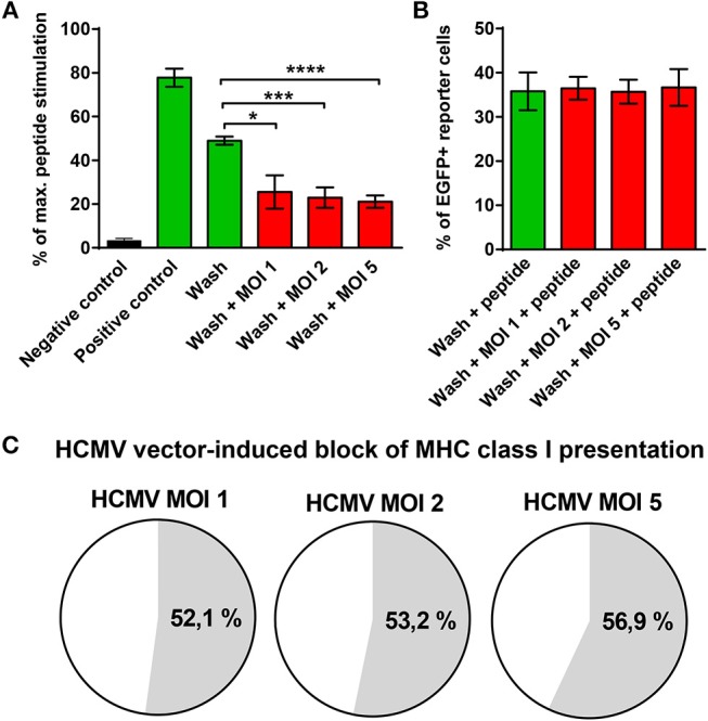
Block of MHC class I presentation induced by immunoevasin-deficient HCMV. U251 cells stably expressing the E6/E7 fusion protein (U251-E6/E7 cells) were left uninfected or infected with RVTB40ΔUS11, a mutant HCMV lacking all known immunoevasins, at the indicated MOIs for 3–24 h. Subsequently, cells were harvested, washed with ice-cold citric acid, to remove all preexisting peptide-MCH complexes and cocultured at a ratio of 2:1 with HPV E6-specific reporter cells, in which NF-κB drives EGFP. After 18 h EGFP expression was assessed by FACS analysis. Unwashed U251 cells (Negative control) and unwashed U251-E6/E7 cells (Positive control) were also cocultured with HPV E6-specific reporter cells. In parallel, maximal peptide stimulation was determined for each experimental group by pulsing cells additionally with E6 peptide (1 μg/ml) before coculture with HPV E6-specific reporter cells and subsequent FACs analysis. (A) The stimulation in each experimental group is given as percentage of maximal peptide stimulation. (B) The % of EGFP+ reporter cells after pulsing with E6 peptide (maximal peptide stimulation) is shown for washed U251-E6/E7 cells left uninfected and washed U251-E6/E7 cells infected with mutant HCMV at the indicated MOIs. (C) The block of MHC class I presentation after infection with mutant HCMV at the indicated MOIs is given as a percentage. The results shown are derived from three independent experiments. Error bars represent the mean ± SEM (****P < 0.0001; ***P < 0.001; *P < 0.05; unpaired t-test).
