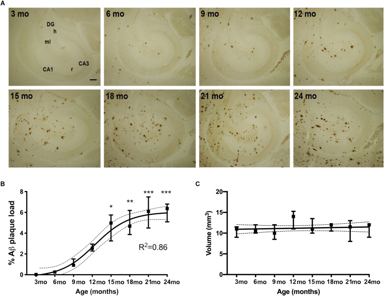
FIGURE 1.
% Aβ plaque load follows a sigmoidal trajectory with age in the hippocampus of APPswe/PS1ΔE9 Tg mice. (A) IHC staining for Aβ shows an age-dependent increase in Aβ plaques in the hippocampus of Tg mice. Initially plaques become abundant in the perforant pathway innervated parts or the hippocampus. CA1, CA3, regio superior and inferior hippocampus, respectively; DG, dentate gyrus; h, hilus; ml, molecular layer; r, stratum radiatum. Scale bar: 100 μm. (B,C) Stereological quantification showing that hippocampal % Aβ plaque load increases significantly from 15 months of age and follows a sigmoidal trajectory, based on non-linear regression analyses (B), whereas hippocampal volume is not significantly changed with age in Tg mice (C). 95% confidence intervals are included. Data points show medians and 25 and 75% quartiles for each group [n = 6/group except for n = 4 for 15-month-old mice in (B), and n = 5 or 6/group except for n = 4 for 15-month-old mice and n = 3 for 24-month-old mice in (C)]. Asterisks represent statistically significant increases in % Aβ plaque load versus 3-month-old mice as determined by Dunn’s test. *p < 0.05, ∗∗p < 0.01, ∗∗∗p < 0.001.

