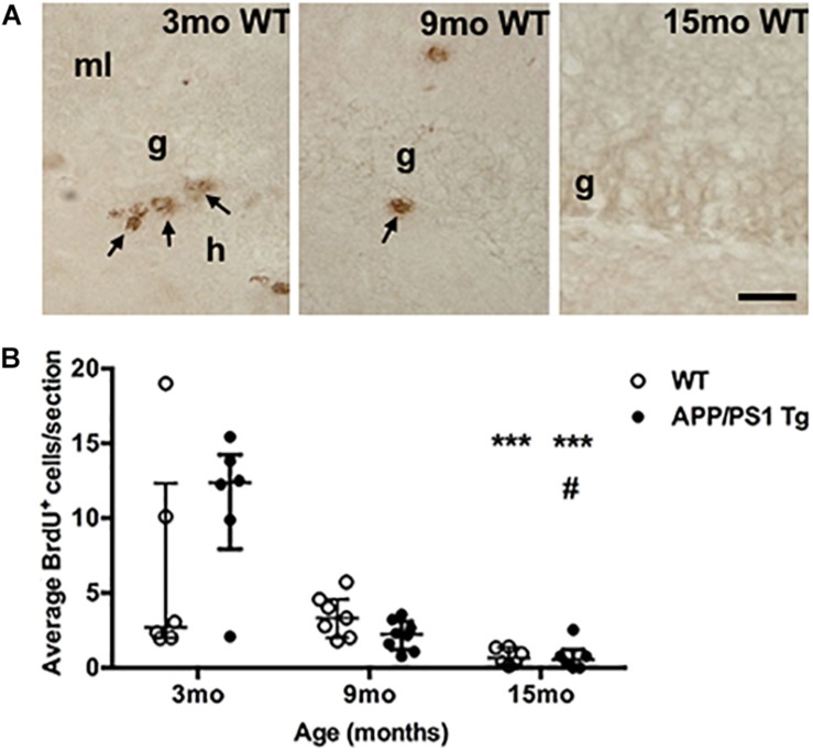
FIGURE 7.
Cell proliferation in the subgranular zone of both genotypes decreases with age. (A) Immunohistochemical staining of proliferating BrdU+ cells (arrows) in the subgranular zone of 3-, 9-, and 15-month-old WT mice. The BrdU-staining was performed on sections from post-fixed, isolated hippocampi. In general, fewer BrdU+ cells were observed in the subgranular zone at 9 months of age, and at 15 months, almost no BrdU+ cells were observed in either Tg (not shown), or WT mice. g, granular cell layer; h, hilus; and ml, molecular layer. Scale bar: 20 μm. (B) Scatter-plot showing the average number of BrdU+ cells in the sgz 3-, 9-, and 15-month-old WT and APPswe/PS1ΔE9 Tg mice. Despite of a large variation in the number of BrdU+ cells in 3-month-old mice (see text for details) there was a significant age-dependent reduction in BrdU+ cells in both APPswe/PS1ΔE9 Tg and WT mice by 15 months of age. Data are expressed as the mean number of BrdU+ cells per section and are shown as mean ± sem for each group (n = 6–8 per group). No significant differences were observed between WT and Tg mice. #p < 0.05 (vs. 9-month-old Tg mice), ∗∗∗p < 0.0001 (vs. 3-month-old mice).

