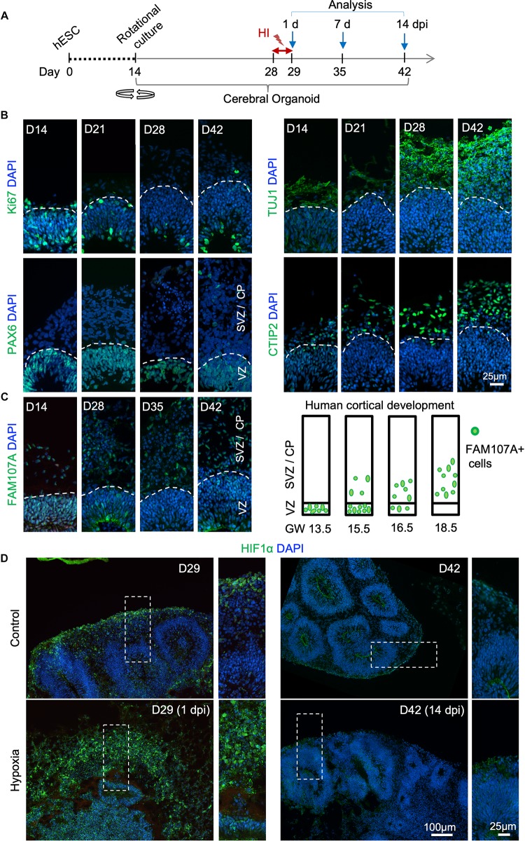
FIGURE 1.
Transient hypoxia model in human cerebral organoids. (A) Schematic diagram of transient hypoxic injury (HI) model with hESC-derived cerebral organoids. D0 represents the starting day of differentiation, D14 denotes the time when organoids were transferred to culture on orbital shaker. At D28, organoids were subjected to 3% O2 in a hypoxic chamber for 24 h, followed by return to 21% O2 culture condition for the remaining culture periods until analysis. (B) IF images for the indicated markers in cerebral organoids demonstrate progressive development of cortex-like structures and appearance of immature neurons (TUJ1+) and deep-layer cortical neurons (CTIP2+) from D14–D42. Proliferating (Ki67+) NSCs (PAX6+) reside in VZ-like germinal region, and offspring in various stages of differentiation migrate out of VZ to populate outer layers, designated as SVZ/CP-like structures. White dashed lines delineate border between VZ and SVZ/CP. Note higher cellular density in the VZ as visualized by DAPI nuclear counterstaining. (C) Left, IF images demonstrate progressive development of oRGs that express FAM107A. Right, schematic depiction of spatial localization of FAM107A+ oRGs during human brain development at the indicated gestational week (GW), adapted from Pollen et al. (2015). (D) Representative IF images show stabilization of HIF-1α at D29 (1 dpi) after hypoxia episode as compared to control, while only baseline HIF-1α immunofluorescence was detected at D42 (14 dpi). High magnification images of boxed areas are shown on the right.

