FIGURE 2.
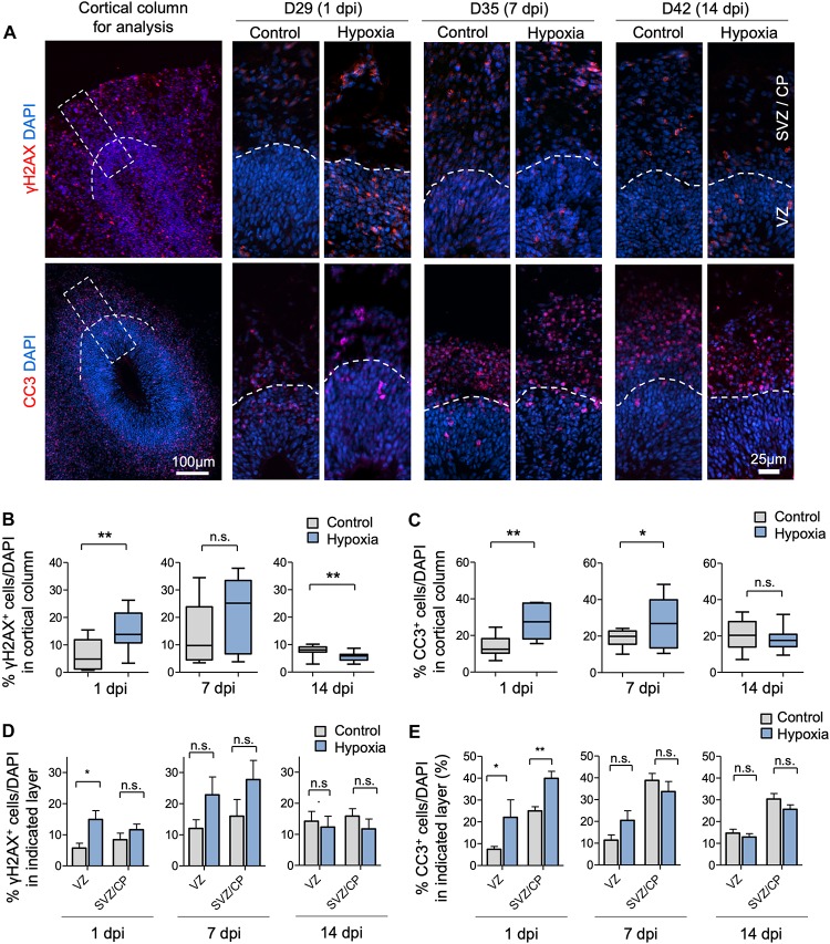
Transient hypoxia induces immediate DNA damage and prolonged apoptosis in cerebral organoids. (A) Representative IF images for γH2AX and cleaved caspase 3 (CC3) in cerebral organoids. Boxed area in lower magnification images in left panel denotes the analyzed area, i.e., a 100 μm wide × 300 μm high radial column spanning all cortical layers (VZ, SVZ, and CP). Corresponding cortical areas of the same size were used for all quantifications in our studies to ensure consistency. Higher magnification images are shown in right panels. White dashed lines demarcate VZ and SVZ/CP. (B,C) Quantifications show the percentage of DAPI+ cells that are positive for γH2AX (B) or CC3 (C). Analyses were carried out in 100 μm × 300 μm cortical column as shown in (A). Student’s t test, n = 9 independent organoids from 3 different batches. (D,E) Quantifications of percentage of DAPI+ cells that are positive for γH2AX (D) or CC3 (E) in VZ or outer layers (SVZ/CP). Two-way ANOVA followed by a Tukey post hoc test; n = 9. *p < 0.05; ∗∗p < 0.01; n.s., not significant.

