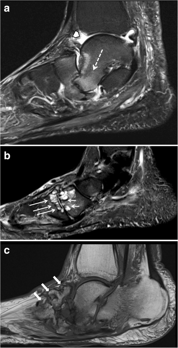
Fig. 17.

Three sagittal images of different patients showing classic features of late-stage Charcot foot. a (Sagittal STIR) inferior dislocation of the talar head (white arrow), effusion in the tibiotalar joint (white arrow head). b (Sagittal STIR) prominent subchondral cysts at the Lisfranc’s joint (white arrows). c (Sagittal T1) bone proliferation and debris in the midfoot (white arrows) and fragmentation of navicular bone
