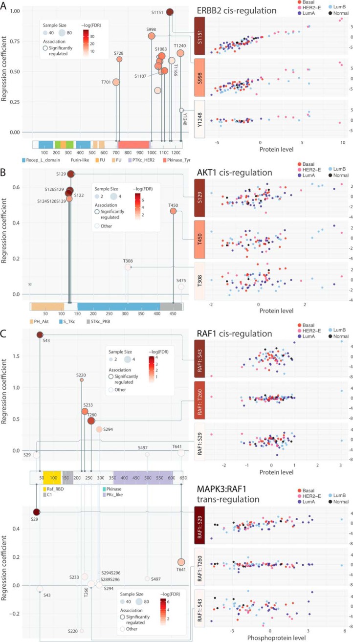
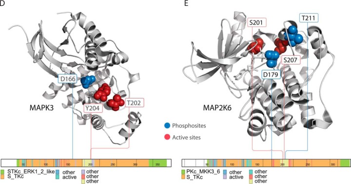
Fig. 4.
Patterns of associated phosphosites on primary sequences and 3D structures. A, Consistent cis-associations of phosphosites identified in ERBB2. B, Discordant cis-associations of phosphosites identified in AKT1. (C) Cis and trans-associations by MAPK3 of RAF1 phosphosites. D, Cis-associated phosphosites p.T202 and p.Y204 in spatial proximity adjacent to the active site p.D166 of MAPK3 as in PDB structure 4QTB (67). E, Trans-associated phosphosites p.S201 and p.S207 (by MAP3K5) are found in spatial proximity to the active sites, including p.D179 and p.T211, of MAP2K6, which is co-crystalized with an ATP analog as in PDB structure 3VN9(68).


