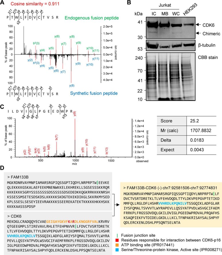
Fig. 5.
Experimental validation and characterization of the FAM133B:CDK6 fusion protein. A, Validation of the identified fusion junction peptide (PTW:LFDVCTVSR) of the FAM133B:CDK6 fusion protein. The m/z value of the matched peaks of the synthetic peptide, which was synthesized based on the identified fusion junction sequence, is consistent with the m/z value of the original matched peaks of the endogenous fusion peptide. B, Dual position of CDK6 detected by antibody-based assay in the Jurkat and HEK293 cells. Fifty micrograms of each fraction of the Jurkat and HEK293 cell lysates were loaded onto a gel, and CDK6 (top panel) and β-tubulin as an intracellular marker (middle panel) were detected by Western blot analysis. The preparative gel was stained using CBB as a loading control (bottom panel). C, The spectrum shows the antibody-captured “chimeric” band (33 kDa) identified as the common peptide (ILDVIGLPGEEDWPR) of FAM133B:CDK6 and wildtype CDK6 by LTQ-Orbitrap-MS. D, The sequences of FAM133B, CDK6, and FAM133B:CDK6 fusion protein with domains from InterPro (https://www.ebi.ac.uk/interpro/) and p16-binding residues are shown. *WC, whole cell lysates; MB, membrane fraction; IC, intracellular fraction; CBB, Coomassie brilliant blue; Mr (calc), relative molecular mass of the matched sequence.

