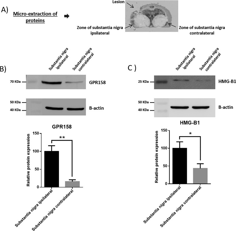
Fig. 3.
Western blotting analyses of proteins extracts using liquid microjunnction extraction collected in ipsilateral and contralateral S. nigra area. A, Picture of a slides on which the liquid microjunction microextraction was performed. B, Western blotting analyses performed with the collected samples with the anti-GPR158 (n = 3) and C, Western blotting analyses performed with the collected samples with the anti-HMGB1 (n = 3).

