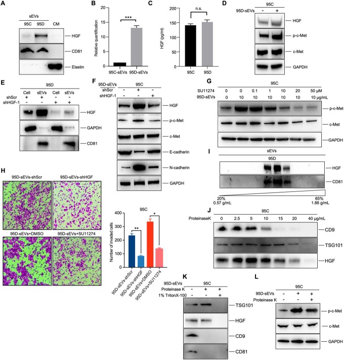
Fig. 5.
sEVs-HGF actives c-Met signaling in recipient cells. A, B, Western blot of HGF protein level in 95C-sEVs and 95D-sEVs. Elastin was used as the negative control for intercellular matrix. Histogram shows the corresponding quantification data. C, ELISA assay of HGF in the culture media from 95C and 95D cells. D, Western blot of c-Met and p-c-Met in recipient cells treated with 10 μg/mL 95D-sEVs or PBS for 3 h. E, Effect of HGF knockdown in 95D-sEVs and cells measured by western blot. F, Target protein expression of 95C cells treated with 10 μg/mL 95D-sEVs-shScr, 10 μg/mL 95D-sEVs-shHGF, or blank PBS for 3 h. G, The phosphorylation of c-Met in 95C cells treated with 95D-sEVs and inhibitor SU11274 were detected by western blot. H, Migration assays of 95C cells treated with 10 μg/mL 95D-sEVs-shScr, 10 μg/mL 95D-sEVs-shHGF, 10 μg/mL 95D-sEVs +DMSO, or 10 μg/mL 95D-sEVs + 20 μM SU11274. The number of migrated cells shows in the right histogram. I, Western blot of HGF protein level in the sEVs, which were purified by sucrose density gradient centrifugation. J, Western blot of HGF and indicated protein markers in 95D-sEVs treated with proteinase K for 30 min at 4 °C. K, Western blot of HGF and indicated protein markers in 95D-sEVs treated with proteinase K for 30 min at 4 °C with or without 1% Triton X-100. L, The phosphorylation of c-Met in 95C cells treated with 10 μg/mL 95D-sEVs or 10 μg/mL protease-treated 95D-sEVs were detected by western blot. Each experiment was performed in triplicates and results are presented as mean ± S.D. (*p < 0.05; **p < 0.01; ***p < 0.

