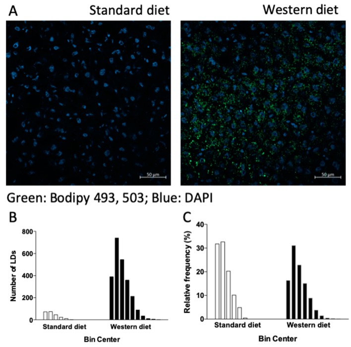
Figure 2.
Lipid droplets (LDs) in rat livers. Staining with Bodipy 493/503, a marker of lipid droplets. Cell nuclei are stained blue with 4′,6-diamidino-2-phenylindole (DAPI). (A). Absolute number of the different classes of LDs according to their diameter (observed range: 0.4–2.4 μm and 0.4–4 μm for rats fed with standard and Western diet, respectively) (B), and their relative frequencies (C).

