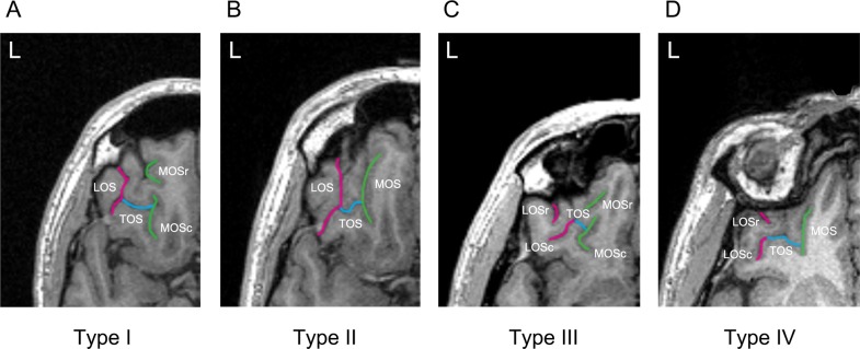
Fig. 1. Classification of the orbitofrontal cortex sulcogyral patterns with magnetic resonance imaging.
Examples of the four major sulcogyral patterns from four different participants. Patterns were classified into four subtypes (Types I–IV) according to the continuity of the lateral and medial orbital sulci (LOS and MOS, respectively) in the rostrocaudal direction (r rostral, c caudal). Type I refers to continuous LOS and discontinuous MOS (a), Type II refers to continuous LOS and MOS (b), Type III refers to discontinuous LOS and MOS (c), and Type IV refers to continuous MOS and discontinuous LOS (d). Sulcal continuities of the MOS and LOS were determined by evaluating several consecutive axial slices rather than just a single slice. TOS transverse orbital sulcus

