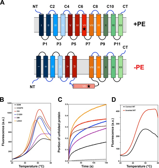
Figure 1.
Inverted LacY stability in various detergents as quantified by the CPM dye binding assay. (A) Topological organization of LacY purified from PE-containing and PE-lacking cells. Cytoplasm is at the top and cylinders indicate TMDs numbered sequentially in Roman numerals from the N-terminal (NT) to the C-terminal (CT) domain. EMDs connecting the TMDs are numbered sequentially and prefixes C and P stand for cytoplasmic and periplasmic, respectively. The star symbol depicts the position of single cysteine replacement I230C in TMD VII. (B) Representative melting curves and (C) representative unfolding curves at 45 °C over the course of 150 min for WT LacY purified from PE-lacking cells and solubilized in 3xCMC of various detergents prior to the assay. LacY was diluted to 26 μg/mL in 50 mM Tris-HCl (pH 7.5), 100 mM NaCl containing 0.05% of the following detergents: CHAPS, OG, LDAO, C12E9, DM and DDM. (D) Melting curves for WT LacY purified from PE-containing (correct topology) and PE-lacking (inverted topology) cells. LacY was diluted to 50 μg/mL in 50 mM Tris-HCl (pH 7.5), 100 mM NaCl containing 0.05% DDM.

