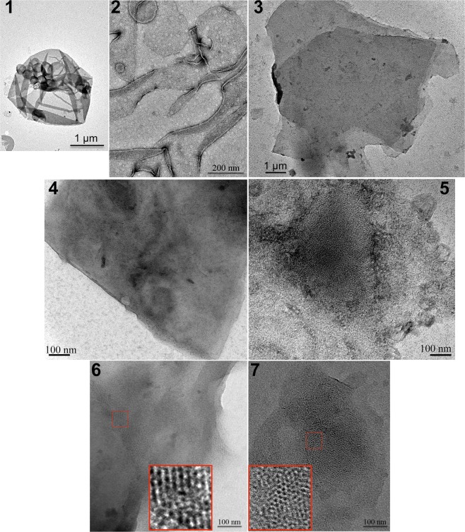
Figure 7.
Electron microscopy of representative crystallization outcomes of the inverted LacY C154G/H205C. Overview of a negatively stained sample depicting the presence of protein reconstitution in densely packed proteoliposomes (panel 1), membrane sheets (panel 2), membrane stacks (panel 3), and patchy crystals (panels 4 to 7). The inverted LacY is visible in patchy crystals areas imaged at higher magnification (panels 6 and 7). Scale bars represent 1 μm in (1) and (3), 200 nm in (2), and 100 nm in (4 to 7).

