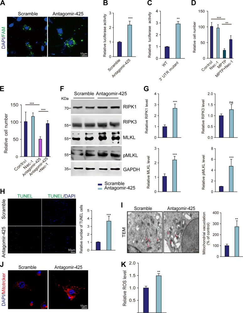
Fig. 3. miR-425 promoted necroptosis by targeting RIPK1.
a FAM immunofluorescence tracing of transfected AntagomiR-425 and scrambled control. b Luciferase activity of PC12 cells cotransfected with the WT 3′UTR of RIPK1 luciferase reporter plasmids together with AntagomiR-425 and scramble control. c Luciferase activity of PC12 cells cotransfected with the WT or mutant 3′UTR of RIPK1 luciferase reporter plasmids together with AntagomiR-425. d Quantification of PC12 cells 3 days after treatment with MPTP, Nec-1, or vehicle control. e Quantification of PC12 cells 3 days after treatment with AntagomiR-425, Nec-1, or vehicle control. f Immunoblotting of RIPK1, RIPK3, MLKL, and pMLKL expression in PC12 cells transfected with AntagomiR-425 or scrambled control. g Quantification of RIPK1, RIPK3, MLKL, and pMLKL expression in PC12 cells transfected with AntagomiR-425 or scrambled control. h TUNEL assay of PC12 cells 3 days after treatment with AntagomiR-425. i Representative mitochondria are shown using TEM and quantification of mitochondria vacuolation. j Representative mitochondria are shown using MitoTracker Red staining. k ROS assay of PC12 cells 3 days after treatment with AntagomiR-425. All data represent the mean ± SEM. In d, e, one-way ANOVA followed by Dunnett’s test was applied. Other experiments used Student’s t test, **P < 0.01, ***P < 0.001, and ns, not significant

