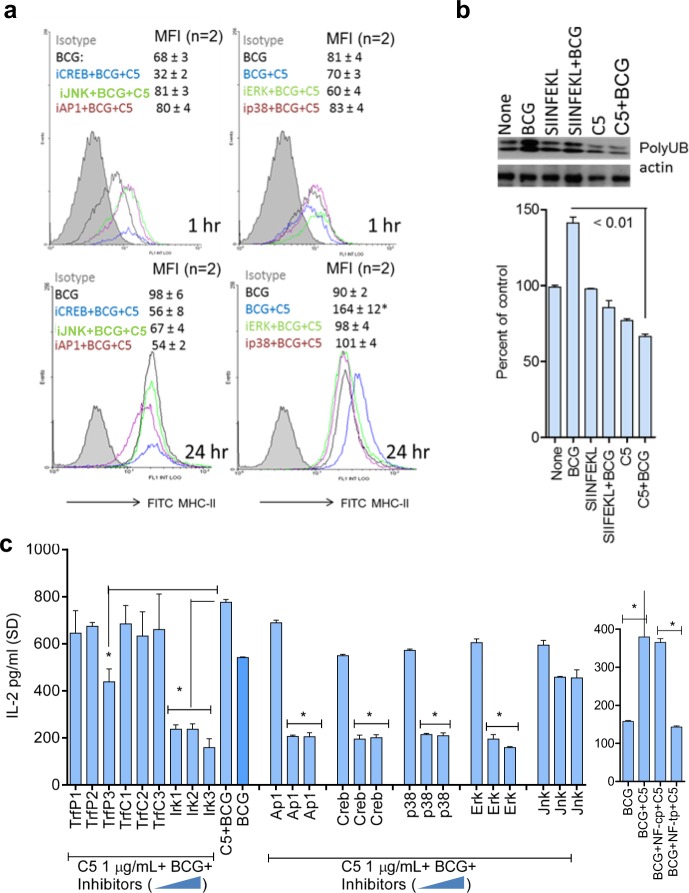
Fig. 4.
CFP-10-derived C5 peptide induces an upregulation of MHC-II in MΦs. a MΦs from wild-type C57Bl/6 mice were tested for surface expression of MHC-II. They were treated or not with 5 µM of inhibitors of API/CREB and MAPK, followed by 2 h activation with C5 peptide, and 1 or 24 h infection with BCG, followed by MHC-II staining and flow cytometry. Inset numbers indicate mean fluorescence intensity (MFI) values averaged from two experiments ( ± SD). C5 peptide enhances MHC-II expression in BCG-infected APCs and blockade of MAPK, AP-1/CREB reduces the levels of MHC-II (*p < 0.01; BCG + C5 group vs. inhibitors). b MΦs were tested naive or activated with C5 (2 h) and BCG (90 min) or irrelevant control SIINFEKL ova-peptide followed by BCG. Cell lysates prepared 4 h later were immune-precipitated with an antibody to MHC-II followed by probing with an antibody for ubiquitinated MHC-II. Densitometry (shown below) indicates that C5 activation decreases the levels of ubiquitinated MHC-II in MΦs, which is increased by BCG infection (two experiments ± SD; t test). c MΦs were incubated for 2 h with increasing doses of (0.5, 1, and 2 µM = lanes 1, 2, 3) specific (TrfP) or control (TrfP) peptide inhibitors of TRAF1/6; a peptide inhibitor of IRAK1/4 (IR1, 2, 3); peptide inhibitors of MAPKs; AP-1 and CREB and NF-kB (NF-tp, specific inhibitor; NF-cp, control peptide), followed by BCG or BCG + C5 for 2 h. Washed monolayers were overlaid with Ag85B-specific CD4 BB7 T cells, and IL-2 in the supernatant was measured after 18 h. Signaling blockade reduces antigen presentation by infected MΦs (*p < 0.009 vs. BCG alone; one-way ANOVA with Dunnett’s post test; one of two similar experiments shown)

