Fig. 4.
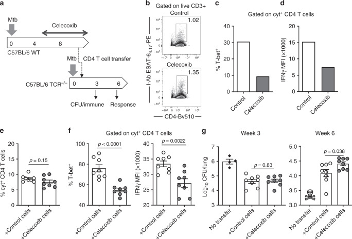
Celecoxib reduce IFNγ expression and diminish the protective capacity of CD4 T cells. a WT C57BL/6 donor mice were infected with ~25–50 CFU Mtb Erdman by the aerosol route and treated for 4 weeks with celecoxib (week 4–8) before purifying CD4 T cells from pooled spleen and tracheobronchial/mediastinal lymph nodes. A total of 9.5 × 10⁶ purified CD4 T cells/mouse were transferred into B6.129S2-Tcratm1Mom recipients (TCR−/−) that were infected 7 days prior to cell transfer. b At the day of transfer, the pool of purified donor cells were characterized for the frequency of antigen specific CD4 T cells by ESAT-64-17 tetramer staining as well as expression of T-bet (c) and IFNγ (d) in CD4 T cells producing any combination of IFNγ, TNFα or IL-2 after stimulation with overlapping ESAT-6 peptides. e Five weeks after cell transfer (week 6 of the infection), the frequency of cytokine producing CD4 T cells in the lungs of the recipient mice was determined by ICS after ESAT-6 peptide stimulation (n = 8). f The cytokine producing CD4 T cells were further characterized based on the expression of T-bet and IFNγ. g Mtb bacilli were enumerated in the recipient animals at week 3 and 6 of the infection by plating lung homogenates (n = 8). All the TCR−/− mice in the control group that did not receive any CD4 T cells met the humane endpoints for euthanasia before week 6. Bars represent mean ± SEM and p values were calculated with an unpaired two-tailed t-test comparing treated and non-treated animals

