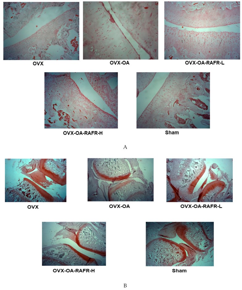
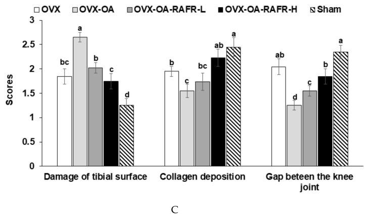
Figure 6.
The histopathological features of osteoarthritic lesions in the knee joints of rats after 21 days of intra-articular injection of monoiodoacetate (MIA) Ovariectomized (OVX) rats were divided into four groups: (1) MIA injection into the knee joint and fed a high-fat diet containing 0.23% rice + 0.018% AFR diet (RAFR-L). (2) MIA injection into the knee joint and fed a high-fat diet containing 0.69% rice + 0.053% AFR diet (RAFR-H). (3) MIA injection into the knee joint and fed a high-fat diet added 0.053% cellulose (OA-control). (4) saline injection into the knee joint and fed a high-fat diet containing 0.053% cellulose (non-OA control). Sham rats had the same diet as the control and received a saline injection (normal-control). At the beginning of the eighth week, an articular injection of monoiodoacetate into the right knee was performed in all OVX groups except the normal-control group, and the assigned diets were provided for an additional three weeks. The depth and extent of cartilage damage and the quantification of the damage were determined in hematoxylin-eosin stained (A), paraffin-embedded knee joint sections from MIA-injected rats (magnifying power ×10). The depth and extent of cartilage damage and quantification of the cartilage damage in the MIA-injected knees were evaluated in Safranin O–fast green–stained (B) knee joint sections from the osteoarthritic rats (magnifying power ×2.5). Scores of the knee joint damage (C) were calculated from the stained sections. Each bar and error bar represents the mean ± SD (n = 5). a,b,c,d Values of the bars with different superscripts were significantly different among groups as per the Tukey test at p < 0.05.


