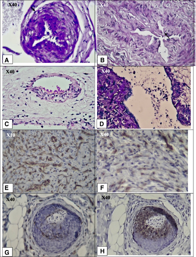
Fig. 2.

Microvascular changes in the patients with diagnosis of thromboangiitis obliterans with long-term medical angiogenic treatment, according to haemotoxylin and eosin (H & E) and Immunohistochemistry (IHC) for CD31 and Ki67. a, b h and e staining. X40 objective lense. Stenosis and shrinkage of the microvessel lumen due to proliferation of endothelial cells. c h and e staining. X40 objective lense The proliferation of endothelial cells can be asymmetrical; thrombus formation is at the site of endothelial cell proliferation. d h and e staining. X40 objective lense. Proliferation of endothelial cells can induce NETosis and further thrombus formation. e IHC for CD31. X10 objective lense. Extensive proliferation of endothelial cells in the soft tissue. f IHC for Ki67. X40 objective lense Mild to moderate positive Ki67 in the soft tissue, which supports the proliferation and mitosis of the endothelial cells.h IHC for CD31. X40 objective lense. Proliferation of endothelial cells in the intima layer. Due to the cut, the lumen of the microvessels cannot quite be seen. Instead, part of the stained endothelial cells is seen. g IHC for Ki67. X40 objective lense. Supporting the mitosis and proliferation of endothelial cells in the intima layer
