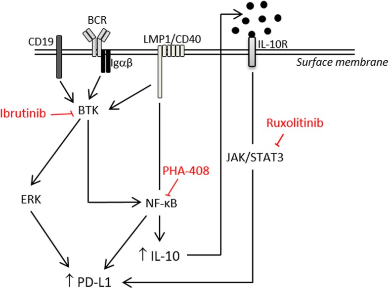
Fig. 5.

Graphical representation of signaling pathways leading to PD-L1 over-expression in tumor B-cells from LMP1/CD40 mice. CD19, BCR (B-cell receptor), Igαβ, LMP1/CD40, and IL-10R (IL-10 receptor) are located at the surface membrane. CD19 plus BCR/Igαβ complex, and CD40 lead to BTK (Burton’s tyrosine kinase) activation which in turn activate ERK and NF-κB that up-regulate PD-L1 expression. Indirectly PD-L1 is also increase by IL-10 by an autocrine loop. PHA-408, inhibitor of NF-κB. Ruxolitinib, inhibitor of JAK1/JAK2. Ibrutinib, inhibitor of BTK
