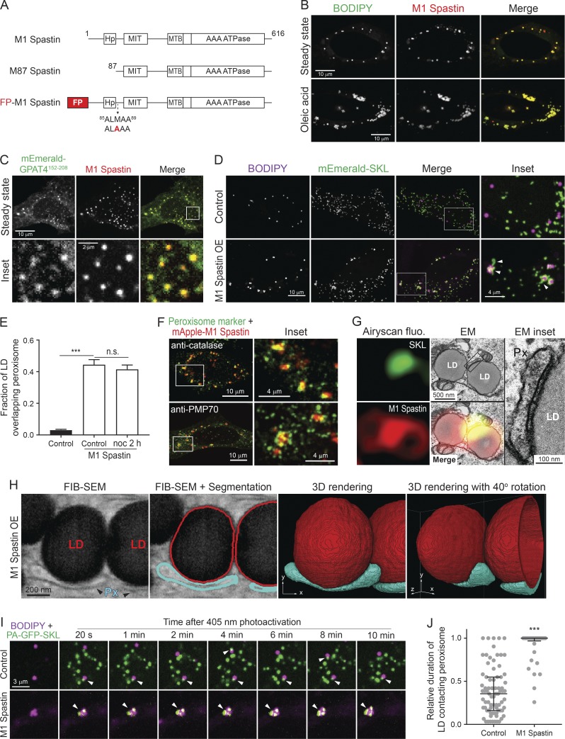
Figure 1.
M1 Spastin promotes LD–peroxisome contact formation. (A) Diagrams of M1 Spastin, M87 Spastin, and FP-tagged M1 Spastin construct. Amino acid number and protein domains are indicated. The mutated residue is labeled in red. (B) mApple-M1 Spastin colocalizes with BODIPY-493/503–labeled LDs in HeLa cells in steady state (top) or following 300 µM oleic acid treatment for 16 h (bottom). Representative confocal images are shown. (C) mApple-M1 Spastin colocalizes with mEmerald-GPAT4152–208 in HeLa cells. Representative confocal images are shown. (D) Association between BODIPY-665/676–labeled LDs and mEmerald-SKL–labeled peroxisomes in control or mApple-M1 Spastin-overexpressing (OE) HeLa cells. Representative confocal MIP images are shown. White arrowheads indicate elongated peroxisomes. (E) Fraction of LD overlapping peroxisome as described in D. mApple-M1 Spastin-expressing cells were further treated with nocodazole (noc). Means ± SEM are shown (22–41 cells from three independent experiments). ***, P < 0.001; n.s., not significant. (F) Association between mApple-M1 Spastin and peroxisomes immunostained with catalase (top) and PMP70 (bottom) antibodies in HeLa cells. Representative confocal MIP images are shown. (G) CLEM images of an LD–peroxisome contact site in mApple-M1 Spastin and mEmerald-SKL expressing HeLa cells treated with 15 µM oleic acid for 16 h. Px, peroxisome. (H) A single 8-nm FIB-SEM slice (left) at LD–peroxisome contacts in mApple-M1 expressing HeLa cells treated with 15 µM oleic acid for 16 h. LDs and peroxisomes (Px) were segmented and reconstructed to reveal LD–peroxisome contacts in 3D. (I) Association between BODIPY-665/676–labeled LDs and peroxisomes labeled by PA-GFP-SKL in control and in mApple-M1 Spastin-overexpressing HeLa cells monitored by confocal microscopy. White arrowheads indicate LD–peroxisome contacts. (J) Relative duration of LD contacting peroxisome as described in I. Median ± interquartile ranges are shown (37–77 LDs from two to three independent experiments). ***, P < 0.001.

