Figure 5.
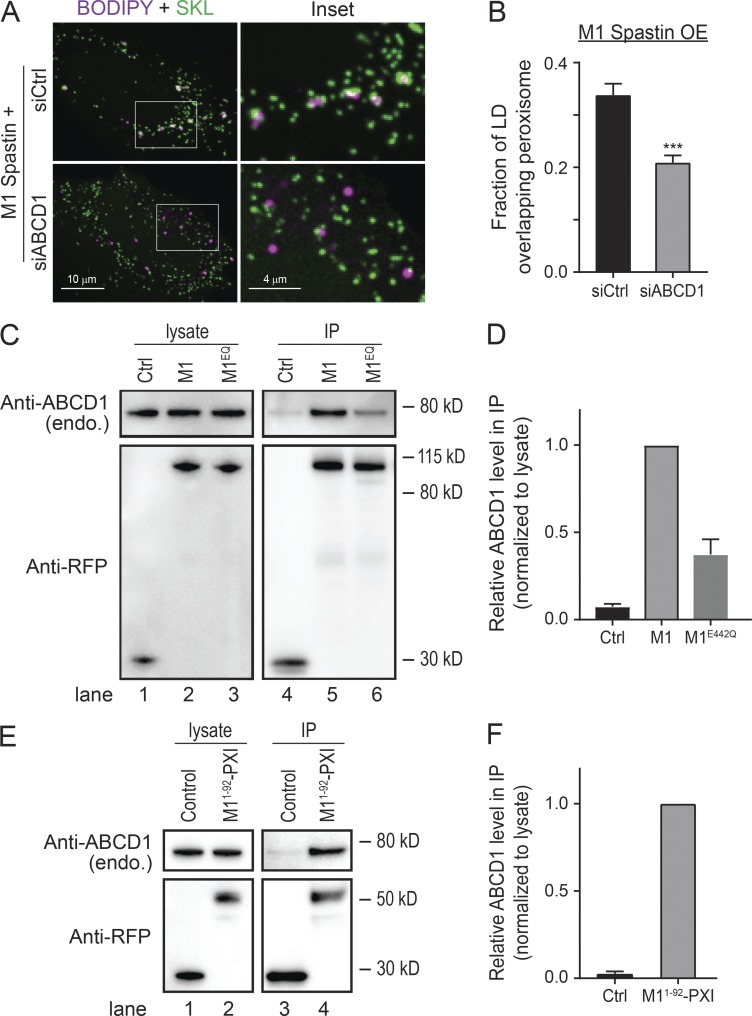
ABCD1 forms a tethering complex with M1 Spastin via the PXI region. (A) Localizations of LDs and peroxisomes in mApple-M1 Spastin-overexpressing (OE) HeLa cells cotransfected with siCtrl or siABCD1. Representative confocal MIP images are shown. (B) Fraction of LD overlapping peroxisome as described in A. Means ± SEM are shown (39–44 cells from four independent experiments). ***, P < 0.001. (C) IP of ABCD1 in HeLa cells transfected with mApple-C1 (Ctrl), mApple-M1 Spastin (M1), or mApple-M1 SpastinE442Q (M1EQ). Protein levels of endogenous ABCD1 and overexpressed constructs were assessed by Western blotting using antibodies against ABCD1 and RFP, respectively. (D) Quantification of relative ABCD1 level in the IP as described in C. The value of M1 is set as 1. Means ± SEM from three independent IP experiments are shown. (E) IP of ABCD1 in HeLa cells transfected with mApple-C1 (Control) and mApple-M11–92-PXI. (F) Quantification of relative ABCD1 level in the IP as described in E. The value of mApple-M11–92-PXI is set as 1. Means ± SEM from two independent IP experiments are shown.

