Figure 5.
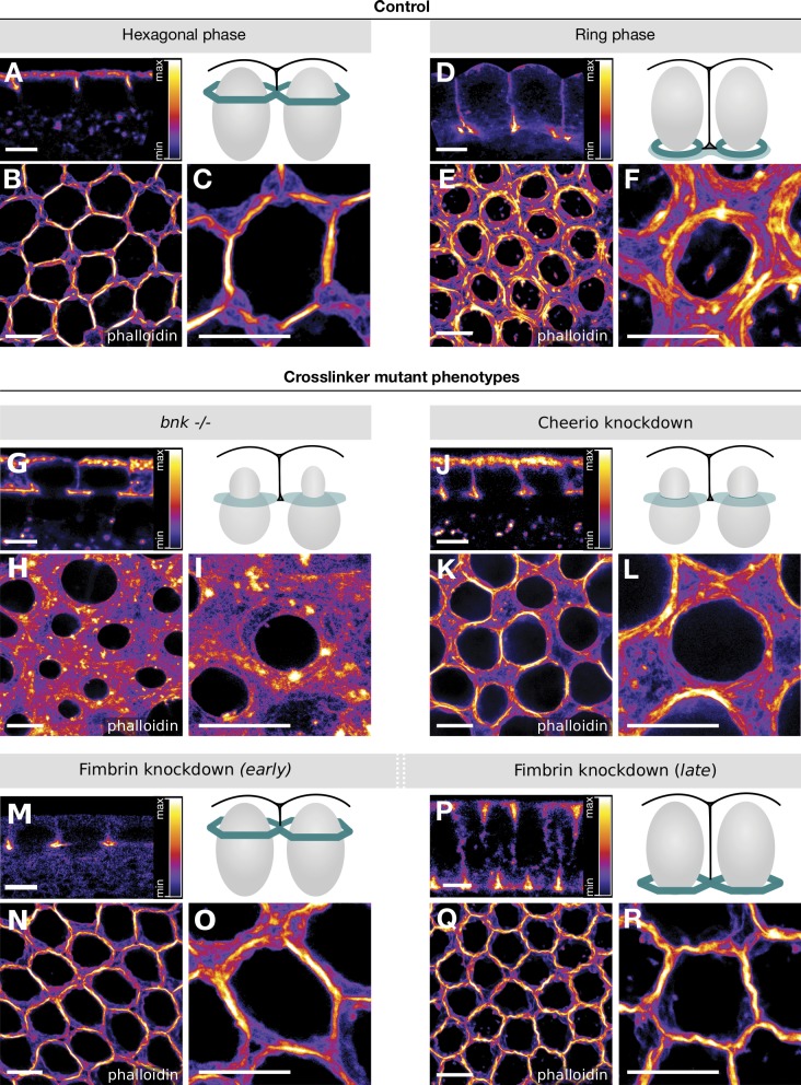
Super-resolution imaging of Cheerio and bnk mutant embryos reveals their critical role in actin bundling during the hexagonal phase. (A–R) Drosophila embryos (wild-type control: A–F; bnk−/−: G–I; Cheerio KD: J–L; Fimbrin KD: M–R) were fixed at different stages of cellularization, stained using phalloidin–Atto 647N, and imaged using 2D-STED microscopy at a resolution of ∼15 nm. Grayscale 8-bit still images were pseudo-colored with the Fire lookup table (LUT, ImageJ software) to produce false-color images. The sagittal cross sections are shown in A, D, G, J, M, and P, the corresponding basal actin networks in B, E, H, K, N, and Q, and closeups (10 µm × 10 µm) in C, F, I, L, O, and R. The data are shown using the Fire lookup table as specified in A, D, G, J, M, and P. Schematics in A, D, G, J, M, and P depict a cross section view of two cells during the corresponding stage of cellularization that was imaged in each respective panel. The basal actomyosin network is colored in green, the nuclei in gray, and the plasma membrane in black. In the wild-type controls (B and C) during the hexagonal phase, actin structures are organized into thick actin fibers. During the ring phase (E and F), actin fibers formed individualized ring structures and a meshwork of actin at the ring interspace. Upon loss of Bnk (G–I), actin fiber formation was severely impaired, and actin was disorganized, forming a meshwork of filaments and thin bundles. KD of Cheerio (J–L) resulted in reduced actin fiber formation, network disorganization, and desynchronized ring constriction. KD of Fimbrin did not affect actin fiber formation during the hexagonal phase (N and O), but rather caused persistence of the actin fiber organized in a hexagonal pattern at later stages of cellularization (Q and R). Scale bars, 5 µm.

