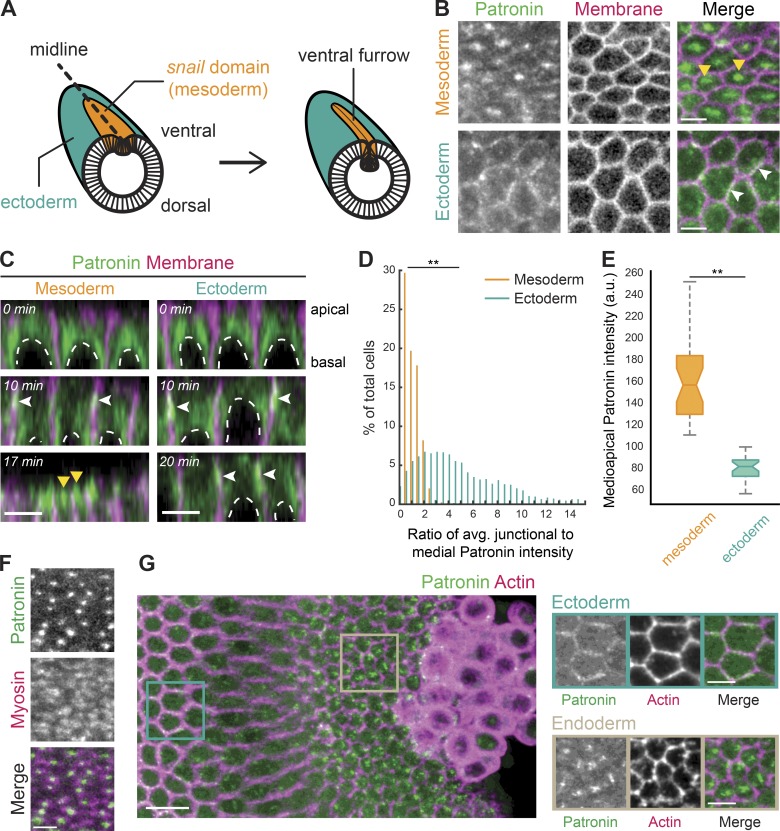
Figure 1.
Patronin::GFP localizes medioapically in apically constricting cells. (A) Diagram of an embryo undergoing mesoderm invagination. Ventral, mesoderm cells (snail expressing domain highlighted in orange) apically constrict and internalize, forming a ventral furrow along the midline (dashed line). (B) Patronin::GFP is present in a medioapical focus specifically in the mesoderm (top row, yellow arrowhead). Patronin::GFP is enriched at junctions in the ectoderm (bottom row, white arrowhead). Images are maximum-intensity projections from a live embryo expressing Patronin::GFP (apical surface) and Gap43::mCH (mCherry-tagged plasma membranes, subapical slice). (C) Patronin::GFP localization changes from junctional (white arrowheads) to medioapical (yellow arrowheads) in the mesoderm. Images are apical–basal cross sections from a live embryo expressing Patronin::GFP and Gap43::mCH. Top row: midcellularization; middle row: late cellularization/early gastrulation; bottom row: during folding. Nuclei are highlighted by dashed white lines. (D) Quantification of medioapical Patronin::GFP enrichment. Individual cells were segmented, the junctional and medioapical Patronin::GFP intensity was calculated, and the distribution of the ratio (junctional/medioapical) was plotted as a percentage of cells within each bin (n = 6 embryos, 559 cells; **, P < 0.0001, Kolmogorov–Smirnov test). (E) Apical Patronin::GFP foci are more intense in the mesoderm than in the ectoderm. The maximum apical Patronin::GFP intensity was determined in a region encompassing the medioapical cortex in both the mesoderm (left) and ectoderm (right; n = 6 embryos, 10 measurements per embryo; **, P < 0.0001, unpaired t test). The notch is the median, bottom and top edges of the box are the 25th and 75th percentiles; whiskers extend to the most extreme data points. (F) Medioapical Patronin::GFP colocalizes with apical myosin patches. Images are apical surface Z-projections from a representative live embryo expressing Patronin::GFP and Myo::mCH (sqh::mCH). (G) Patronin::GFP localizes medioapically in apically constricting endoderm cells. Images are maximum-intensity projections of a fixed embryo expressing Patronin::GFP (apical surface). The embryo was immunostained with Phalloidin conjugated with AF647 to visualize cell outlines (subapical section). Scale bars represent 10 µm (G, left) and 5 µm (B–F and G, right).

