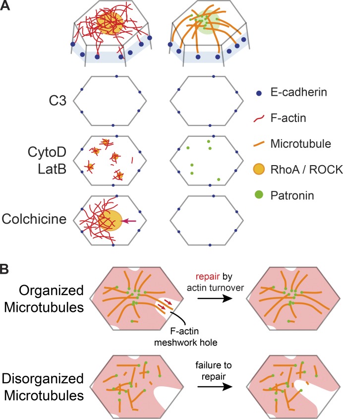
Figure 7.
Actin and microtubule cytoskeletons interact to promote intercellular force transmission. (A) Diagrams at top show the proposed organization of the microtubule and actin cytoskeletal networks at the cell apex. Diagrams below show top down views of the apical cortex (without microtubules) and the effect of various perturbations on Rho/ROCK/F-actin and Patronin localization. C3 injection eliminates apical RhoA/ROCK and Patronin foci. CytoD/LatB injections lead to formation of smaller RhoA/ROCK and Patronin puncta. Colchicine injection does not affect RhoA/ROCK polarity but leads to actomyosin network separations from intercellular junctions (arrow). (B) A model for how microtubules promote reattachment of the apical F-actin meshwork to adherens junctions after fracture. In wild-type embryos (top), the organization of apical, noncentrosomal microtubules could promote repair of holes in the F-actin meshwork (red) by guiding F-actin polymerization and/or bundling through physical associations (red arrows). When microtubule organization is disrupted (bottom), the hole in the F-actin meshwork cannot be repaired in a timely manner, leading to actomyosin network separation from adherens junctions.

