Figure 5.
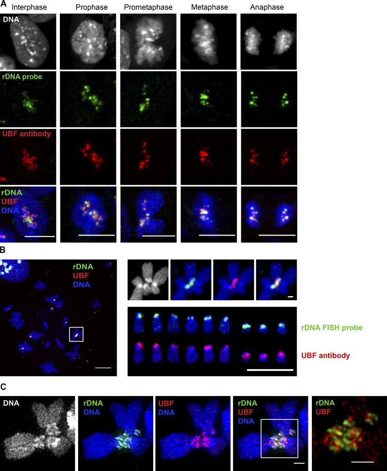
UBF is associated with rDNA loci throughout mitotic progression and with rDNA linkages. (A) Localization of UBF and rDNA during a progressive sequence of mitotic stages. Immuno-FISH of fixed RPE1 cells labeled with rDNA FISH probe (green) and UBF antibody (red). UBF is associated with rDNA in interphase and throughout mitotic progression. Bar, 10 µm. (B) A chromosomal spread from an RPE1 cell labeled by immuno-FISH with rDNA probe (green) and UBF antibody (red). The white box on the left shows the rDNA linkage shown separately on the right (top). Both rDNA and UBF form a bridge between two chromosomes. The panel on the lower right shows individual acrocentric chromosomes labeled with rDNA probe and UBF antibody, respectively. All rDNA loci in this cell line contain UBF. Bar, 10 µm. (C) SIM images of rDNA-linked mitotic chromosomes from c-Myc–overexpressing RPE1 cell line cMyc-3 labeled by immuno-FISH with rDNA probe (green) and UBF antibody (red). Both rDNA and UBF form filamentous connections between chromosomes. Bar, 1 µm.

