Figure 6.
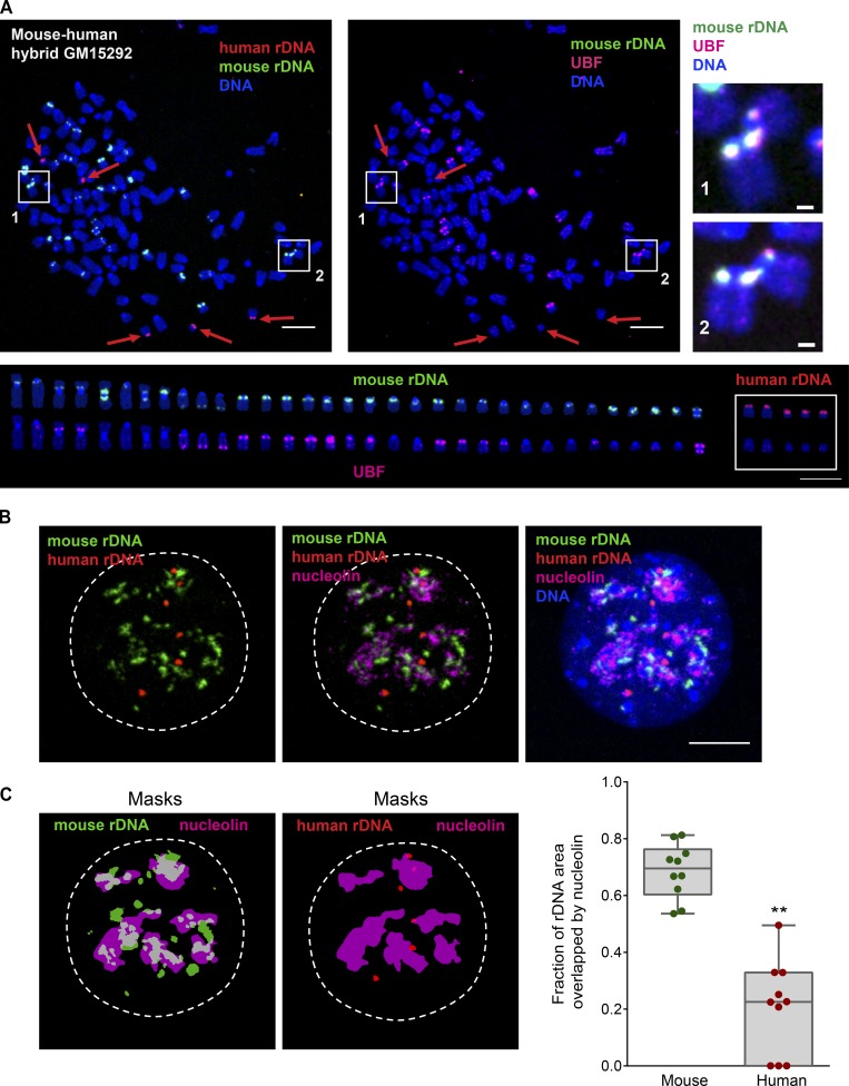
Human–mouse hybrid cell line GM15292 displays rDNA linkages only between active (UBF+) mouse rDNA loci. (A) A representative mitotic chromosome spread from mouse–human hybrid cell line GM15292 labeled by immuno-FISH with mouse rDNA probe (green), human rDNA probe (red), and UBF antibody (magenta). Boxes 1 and 2 highlight rDNA linkages (magnified on the right; bar, 1 µm). Red arrows point to human rDNA chromosomes. The lower panel shows individual mouse and human rDNA chromosomes arranged according to their species and size. The top row shows rDNA probe labeling, and the bottom row shows UBF antibody labeling of the same chromosomes. This particular spread had 112 total chromosomes, of which 33 were mouse rDNA chromosomes (19 UBF+) and five were human rDNA chromosomes. Note that rDNA linkages were formed only between mouse rDNA chromosomes positive for UBF (active). In all human rDNA chromosomes, loci with rDNA were UBF negative (silenced) and did not form linkages. At least 10 chromosomal spreads were examined. Bar, 10 µm. (B) An example of the interphase nucleus from mouse–human hybrid cell line GM15292 labeled by immuno-FISH with mouse rDNA probe (green), human rDNA probe (red), and nucleolin antibody (magenta). While most mouse rDNA is decompacted and associated with nucleolin, human rDNA loci remain compacted and are not incorporated in nucleoli. At least 10 nuclei were examined. Bar, 10 µm. (C) The fraction of rDNA area overlapping with nucleolin in mouse–human hybrid cell line GM15292. The area of overlap was determined between binary masks of mouse rDNA (green) and nucleolin (magenta), and between human rDNA (red) and nucleolin (magenta). Images of masks correspond to the nucleus shown in B. The graph on the right shows fractions of areas overlapping with nucleolin from 10 nuclei. A t test was used to compare nucleolin-overlapping area fractions of mouse and human rDNA. **, P < 0.0001.

