
Figure 2.
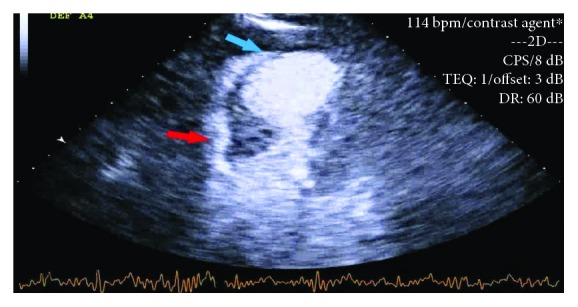
Transthoracic echo showing severely reduced ejection fraction of 30-35%, severe hypokinesis of mid to apical segment with more involvement of the mid anteroseptum, and anterior wall (blue arrow) with basal hyperkinesia and basal asymmetric hypertrophy of the septum (red arrow).