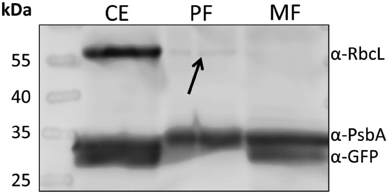
Fig. 4.
Western blot analysis of the final mitochondria (MF) and plastids fraction (PF). 0.5 �g of total protein per lane were tested with three different compartment marker antibodies: α-RbcL, antibody against Rubisco large subunit [marker for plastid stroma]; α-PsbA, antibody against photosystem II protein D1 [marker for thylakoids]; α-GFP, antibody against green fluorescent protein [marker for mitochondrial matrix proteins]. CE, Tig19 cell extract; PF, plastids fraction; MF, mitochondria fraction.

