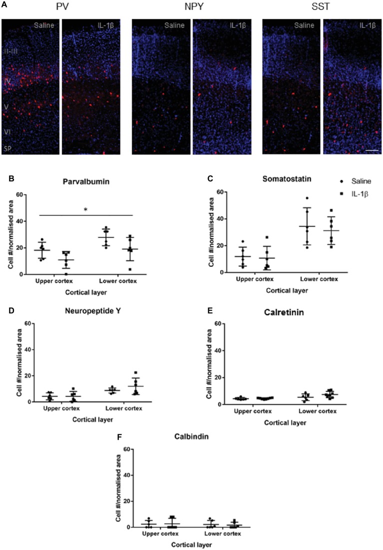
Figure 3.
Developmental pattern of interneuron injury changes by P10 in mice with inflammation-induced brain damage. Interneuron populations were assessed again at P10 by immunohistochemistry (A) and quantified (B–E) for number and distribution through cortical layers. There was a significant decrease in the number of parvalbumin (PV)-positive interneurons across the cortex (B; treatment effect, p = 0.01, two-way ANOVA), but no change for somatostatin (SST), neuropeptide Y (NPY), calretinin (CalR), or calbindin (CalB, C–F). Data presented as mean ± SD; scale bar = 100 μm. *p < 0.05.

