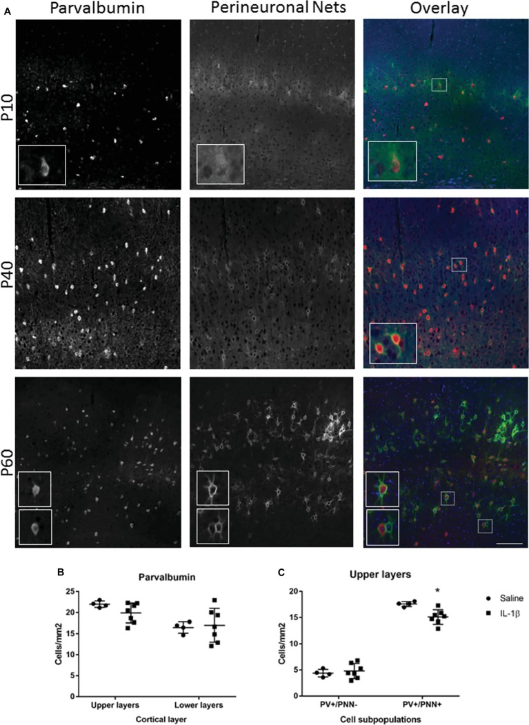
Figure 4.
Long-term changes in parvalbumin-positive interneurons and their perineuronal nets extend to P40 in mice with inflammation-induced brain damage. The beginning of perineuronal net formation could be seen in the cortex, primarily in layer IV, from as early as P10 (A, top row). The aggregation of the perineuronal nets and their association with the parvalbumin-positive interneurons became more pronounced through development (A, lower rows). In the IL-1β challenged mice, there was no gross alteration in perineuronal net formation or in the number of cortical parvalbumin neurons (B). However, there was a significant decrease in the number of PV+/PNN+ neurons in the upper layers of the cortex (C). Data presented as mean ± SD; scale bar = 100 μm. *p < 0.05. PV, parvalbumin); PNN, perineuronal nets.

