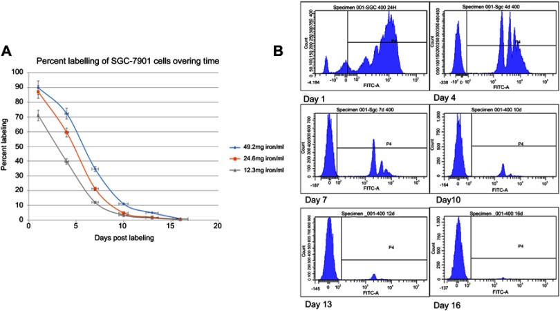
Figure 1.
In vitro dilution of MPIO in SGC-7901 cells that were exposed to a range of particle concentrations as measured by flow cytometry. (A) Flow cytometry data of labeling efficiency for different particle concentrations is shown. (B) Plots of flash green fluorescence intensity show that the average iron content of the SGC-7901 cells reached a plateau (90.0%) after 24 hrs of incubation with MPIO at a concentration of 49.2 mg iron/ml (based on unlabeled control sample).

