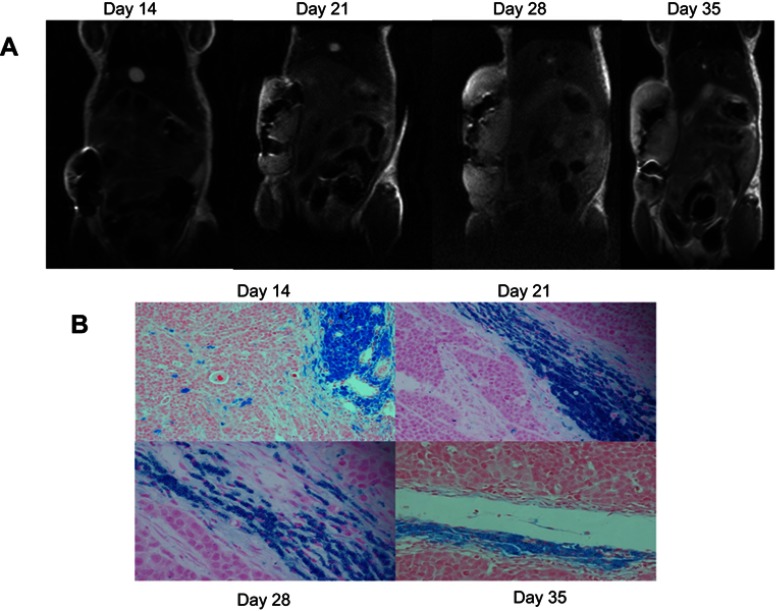
Figure 3.
Coronal in vivo T2-weight images of labeled tumors over time and the corresponding PPB-stained sections. The tumor growth from iron-labeled SGC-7901 cells was monitored postinjection. The signal loss caused by the iron-labeled cells was evident at day 14 postinjection and persisted as the tumor developed. PPB-stained sections show the presence of iron-labeled cells within the tumor. (A) MR image of the labeled tumor. (B) PPB-stained section showing the iron content of labeled tumor over time.

