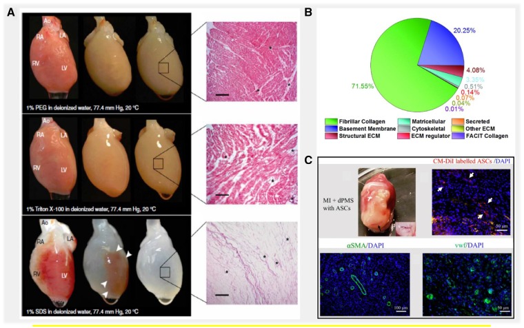
Figure 2.
Advantages of using cardiac tissue-derived decellularized extracellular matrix for myocardial repair. (A) Perfusion decellularization of rat WHs using poly ethylene glycol (PEG), Triton X-100 and SDS. Corresponding H&E staining of dECM showed complete decellularization of rat heart perfused with SDS and incomplete decellularization in PEG and Triton X-100-treated hearts. Scale bar. 200 µm. Source: Adapted with permission from Ott et al. [14]. (B) Measurement of total proteins obtained from injectable human cardiac dECM using ECM-targeted quantitative conCATamers (QconCAT) by liquid chromatography–selected reaction monitoring (LC-SRM). Source: Adapted with permission from Johnson and Hill et al. [37]. (C) In vivo assessment of cardiac dECM generated by seeding adipose-derived stem cells (ASCs) in a rat myocardial infarction (MI) model. Vessel formation was demonstrated as shown by immunofluorescence staining with vascular markers α-SMA and vWF. Source: Adapted with permission from Shah et al. through open access policy [38]

