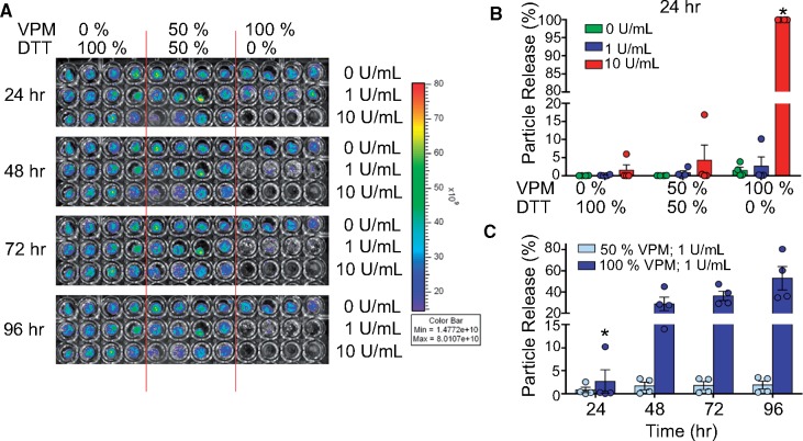
Figure 3.
PEG-4MAL Hydrogels release therapeutics in a formulation and protease-dependent manner. (A) Fluorescent particle release over time imaged by IVIS. (B) Flow cytometry analysis of nanoparticle release from PEG-4MAL hydrogels when exposed to collagenase as a function of protease concentration and crosslinker composition (percentage of DTT vs. VPM). (C) Scatter plot comparing particle release over time of hydrogels fabricated with 50% VPM vs. 100% VPM hydrogels. All groups used n = 4 hydrogels; *P < 0.05 compared to all other groups.

