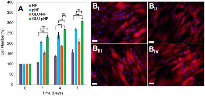
Figure 6.
The Percentage of increase in cell number at 1, 3 and 7 days after seeding cells on neat NF, NTAP treated NF (pNF), GLU peptide conjugated NF (without NTAP treatment) (GLU-NF) and NTAP treated GLU peptide conjugated NF (GLU-pNF). (AI) Morphology of human marrow stromal cells (hMSCs) seeded on NF (BI), pNF (BII), GLU-NF (BIII) and GLU-pNF (BIV). PLGA NF incubated in basal media for 7 days. In the images, cell nuclei and cytoskeletal actin are stained with DAPI (blue) and phalloidin (red) (scale bar represents 50µm)

