Figure 1.
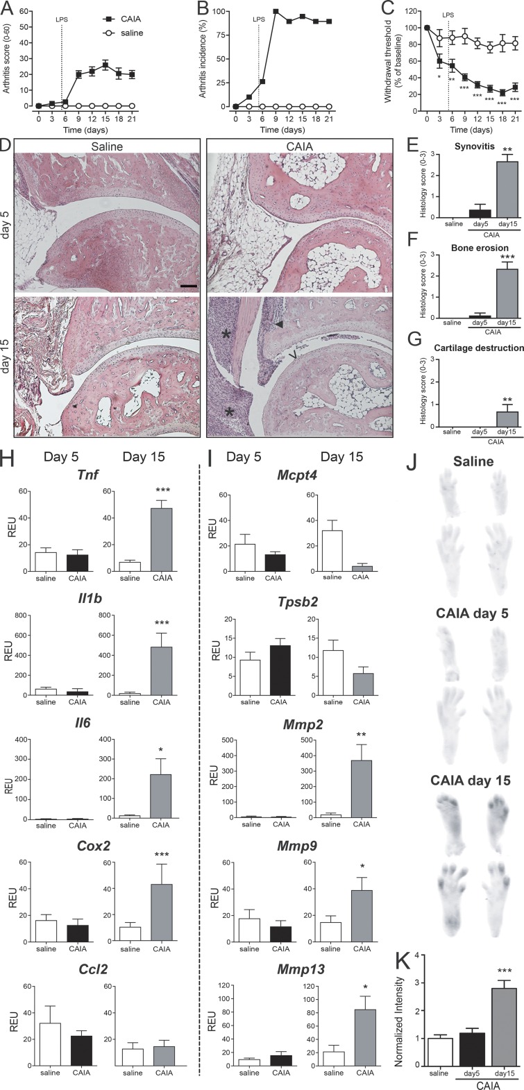
Injection of anti-CII antibodies induces pain-like behavior before visual, histological, and molecular signs of arthritis. (A–C) B10.RIII mice injected with anti-CII mAbs (n = 19; saline controls n = 17) started developing joint inflammation around day 6 (A). On day 9, all animals displayed signs of arthritis (B). Mechanical hypersensitivity (C) was observed already on days 3 and 5, before onset of arthritis, and persisted throughout day 21. (D) Representative H&E histology of B10.RIII mouse ankle joints collected 5 and 15 d after injection of anti-CII mAbs. While an inflammatory infiltrate, bone erosion, and cartilage serration were visible on day 15, no signs of joint pathology was detectable on day 5 or in saline controls. Scale bar represents 100 µm. *, ▼, and V point to signs of synovitis, bone erosion, and cartilage destruction, respectively. (E–G) Scores for inflammatory hallmarks as synovitis (E), bone erosion (F), and loss of cartilage (G) revealed mild ankle joint pathology in two of eight mice day 5 and prominent signs in all mice day 15 (n = 5). Control mice represent pooled time-matched saline-injected mice (n = 4+4). (H and I) Quantitative PCR analysis of joint extracts showed a significant increase in mRNA levels of most of the inflammatory factors investigated at day 15 (n = 7), while none of them were elevated at day 5 of CAIA (n = 6), compared with saline controls (n = 5; H and I). (J and K) Activation of MMPs was significantly increased only after 15 d of CAIA, while no changes were detected at day 5 (n = 3/group, B10.RIII mice). Data are presented as mean ± SEM. *, P < 0.05; **, P < 0.01; ***, P < 0.001 compared with saline controls. REU, relative expression unit.

