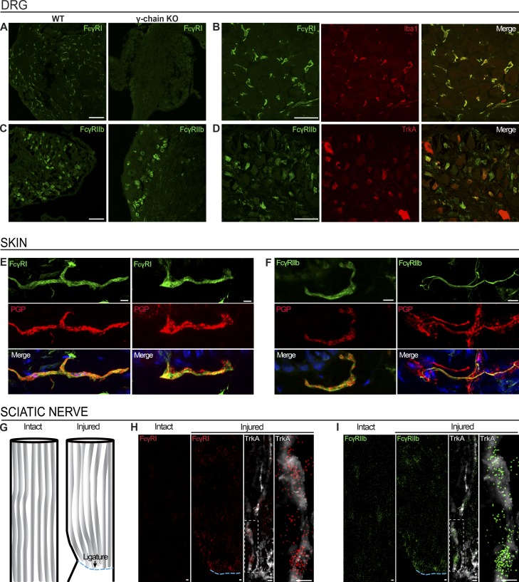
Figure 5.
FcγRI and FcγRIIb are expressed in the DRG and in nerve fibers in the skin. (A and B) FcγRI immunoreactivity was detected in WT BALB/c DRGs, but not in FcRγ-chain−/− mice (A), colocalizing with Iba1-positive resident macrophages (B). Scale bars represent 100 µm and 50 µm, respectively. (C and D) FcγRIIb immunoreactivity was detected in BALB/c mouse DRG and retained in FcRγ-chain−/− mice (C), colocalizing with TrkA-positive neurons (D). Scale bars represent 100 µm and 50 µm, respectively. (E and F) FcγRI (E) and FcγRIIb (F) immunoreactivity was detected in PGP9.5-positive nerve fibers in BALB/c mouse glabrous skin. Scale bars represent 5 µm. (G–I) smFISH on BALB/c mouse sciatic nerves after ligation (G) revealed accumulation of mRNA molecules for Fcgr1 (H) and Fcgr2b (I) proximal to the site of ligation (ipsilateral), while barely any signal was found in the contralateral intact nerve. Scale bars represent 10 µm. See also Figs. S2, S3, and S4.

