Figure 9.
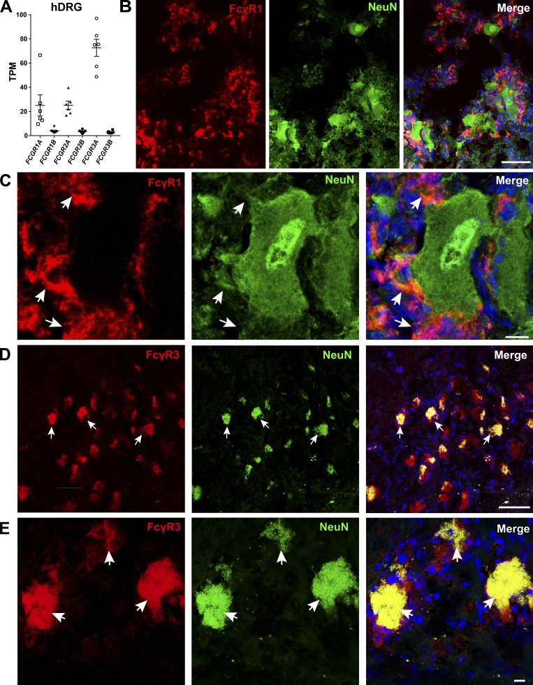
FcγRI and FcγRIII are expressed in human DRG. (A) Publicly available data show the presence of Fcgr mRNA in human DRGs (n = 6). Fcgr3a is the most highly expressed. (B and C) FcγRI immunoreactivity was detected in human DRGs (n = 4). The lack of colocalization with NeuN and the morphology of the FcγRI-positive cells suggest FcγRI expression in resident macrophages, similarly to mice (scale bars represent 100 µm and 10 µm in close-up images). White arrows indicate FcγRI-positive cells, which are negative for NeuN. (D and E) Immunoreactivity for the activating FcγRIII in human DRGs (n = 4) colocalized with the neuronal marker NeuN (scale bars represent 100 µm and 10 µm, respectively). White arrows point to double-positive neurons (FcγRIII and NeuN).

