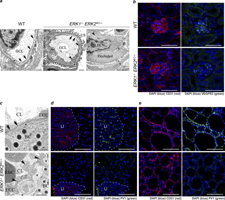
Figure 5.
Loss of endothelial fenestration in glomeruli endothelium and endocrine gland endothelium in Erk1−/− Erk2iEC−/− mice. (a) Transmission electron photomicrographs of kidney Erk1−/− Erk2iEC−/− mice reveal swollen endothelial cells (arrowheads) with loss of glomerular capillary cell fenestrations (arrows) and basement membrane (*) thickening and distortion with localized effacement of podocyte foot processes (p) and scattered occlusion of the glomerular capillary lumen (GCL) by fibrin (presumptive). ECN, endothelial cell nucleus; US, urinary space. Representative images from n = 4 mice per genotype. Scale bars, 500 nm. (b) Immunofluorescent staining of VEGFR2 (green) in glomeruli from Erk1−/− Erk2iEC−/− and WT mice 2 wk after tamoxifen injections. Representative images from n = 4 mice per genotype. Scale bars, 50 µm. (c) Transmission electron photomicrograph of pancreas from Erk1−/− Erk2iEC−/− mice reveal swollen endothelial cells (arrowheads) with loss of capillary endothelial cell fenestrations (arrows) and basement membrane (*) thickening and distortion compared with WT mice. BC, β cells; CL, capillary lumen; ECC, endothelial cell cytoplasm. Representative images from n = 4 mice per genotype. Scale bars, 500 nm. (d and e) Immunofluorescent staining of PV1 (green) in pancreas (d) and thyroid (e) in Erk1−/− Erk2iEC−/− and WT mice. Representative images from n = 4 mice per genotype. Scale bars, 50 µm. LI, Langerhans islet.

