Figure 6.
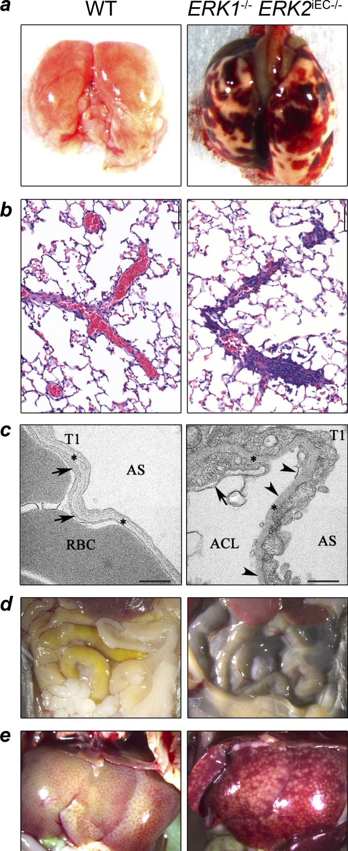
Pulmonary, hepatic, and intestinal hemorrhage in Erk1−/− Erk2iEC−/− mice. (a) Erk1−/− Erk2iEC−/− mice 4 wk after tamoxifen injection show macroscopic pulmonary and pleural hemorrhage compared with WT. (b) Erk1−/− Erk2iEC−/− mice necropsied 28 d after tamoxifen injections have partially or completely occluded pulmonary vessels compared with WT mice (n = 4 mice per genotype; a thrombus was found in three out of four mice). (c) Lung electron photomicrographs from Erk1−/− Erk2iEC−/− mice show capillary endothelial cell membrane (arrows) loss and delamination (arrowheads) compared with WT mice. Arrowheads show the capillary endothelial cell membrane, and asterisks show the basement membrane. RBCs within the alveolar capillary lumen (ACL) are shown. AS, alveolar space; T1, alveolar type 1 epithelial cell. Scale bars, 500 nm. (d and e) Erk1−/− Erk2iEC−/− mice 5 wk after tamoxifen injection show macroscopic dark loops of intestine (d) and hepatic sinusoidal congestion (e) consistent with intraluminal and hepatic hemorrhage, respectively, compared with WT mice.

