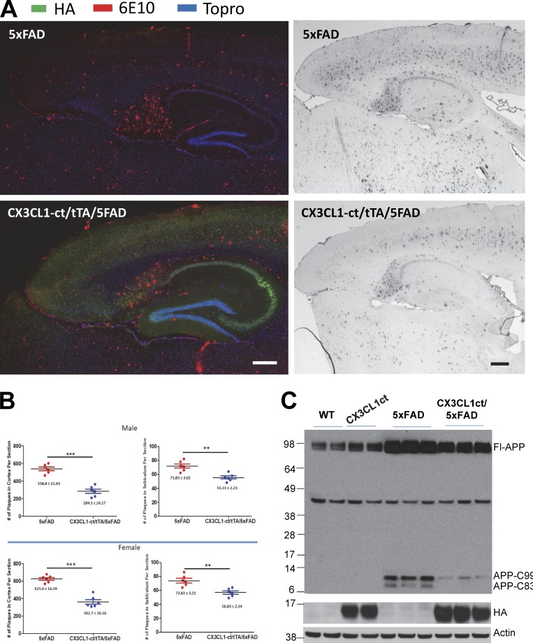
Figure 3.
Overexpression of CX3CL1-ct in 5xFAD transgenic mice reduces amyloid deposition. (A) Two different pairs of Tg-5xFAD and Tg-CX3CL1-ct/tTA/5xFAD mice were used for comparing the density of amyloid plaques by confocal or DAB staining. Antibody 6E10 recognizes human Aβ peptides and amyloid plaques, while HA-tagged CX3CL1-ct transgene was detected with HA antibody. Cell nucleus was marked by Topro. Scale bars, 200 µm. (B) Densities of amyloid plaques from subiculum or cortex were quantified from six mice in each pair. Both male and female mice were used for comparisons. Error bars are ± SEM. (C) APP and its cleavage product C99 and C83 were examined by antibody 8717 (n = 3 experiments). **, P < 0.01; ***, P < 0.001.

