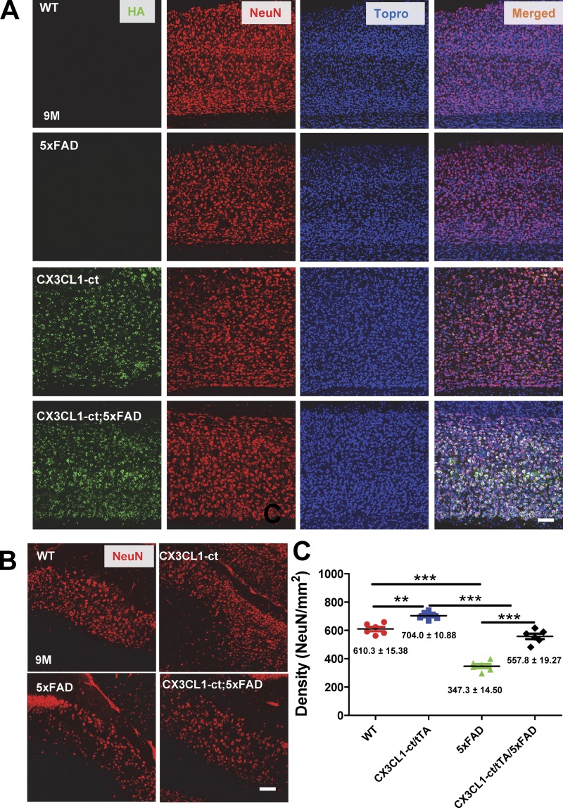
Figure 4.
Ectopic expression of CX3CL1-ct in 5xFAD mice reverses neuronal losses. (A and B) Neuronal loss was observed in the 5xFAD subiculum (A) and cortical layers (B) compared with WT controls. Higher neuronal density, marked by NeuN antibody, was noted in mice overexpressing CX3CL1-ct, suggesting a potential increase in neurogenesis. Ectopic expression of CX3CL1-ct in 5xFAD brains showed more neurons compared with 5xFAD comparable regions. Scale bar, 30 µm. (C) NeuN-marked neurons in the subiculum region were counted for comparison, one in every 10 sections per mouse. Each dot represents one animal (n = 6; **, P < 0.01; ***, P < 0.001, Student’s t test); error bars are ± SEM.

