TABLE 1.
Studies using specifically named defined communities to study host-microbe interactions in vivo (n = 31)a
| Consortium (no. of speciesb) | Division of phylae | Strain source(s) | Host species (strain) | Part of the gut studiedf | No. of animals/group | Chow | Sex | Age (collection time[s]c) | Study outcome(s)d | Reference |
|---|---|---|---|---|---|---|---|---|---|---|
| Schaedler flora (5 species) 2 Lactobacillus spp., anaerobic Streptococcus sp. (group N), Bacteroides strain, Enterococcus sp., coliform strain |
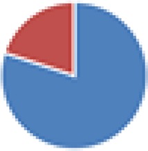
|
Mouse | Mouse (NR) |

|
20 | NR | NR | 4 wk (3 wk–4 mo) | Colonization pattern; cecal size | 9 |
| ASF (8 species) ASF356: Clostridium species ASF360: Lactobacillus intestinalis or Lactobacillus acidophilus ASF361: Lactobacillus murinus or Lactobacillus salivarius ASF457: Mucispirillum schaedleri ASF492: Eubacterium plexicaudatum ASF500: Pseudoflavonifractor sp. ASF502: Clostridium sp. ASF519: Parabacteroides distasonis |

|
Mouse | Mouse (HA/ICR) |

|
30 | NR | Both | Adult (14–56 days) | Death after C. botulinum infection; fecal C. botulinum toxin excretion; colonization pattern of C. botulinum | 36 |
| Mouse | Rat (F344) |

|
1–5 | Sterile food (Charles River) ad libitum | M | NR (2 wk) | Hepatic genotoxicity of mononitrotoluene isomers; metabolic activation of 2NT by intestinal bacteria; cecal bacterial content | 169 | ||
| Mouse | Mouse (scid C.B-17) |

|
4–6 | Autoclaved pelleted diet ad libitum | NR | NR (8–12 wk postreconstitution CD4+ T cells) | After Helicobacter hepaticus infection, rectal prolapse; clinically severe disease; grossly thickened colon, cecum, and rectum on necropsy; colonic inflammation score; colonic epithelial cell proliferation; histopathology | 16 | ||
| Mouse | Rat (HLA-B27 on 33-3/F344) |

|
7–11 | NR | At least M | 2 mo (1 mo) | Gross gut score, levels of MPO and IL-1B in cecal tissue; histological inflammatory score of cecum and antrum | 15 | ||
| Mouse | Mouse (C3H/HeN) |

|
4–8 | Irradiated diet (Harlan Teklad) | NR | 6–8 wk (9–14 wk) | After colonization with Helicobacter bilis or Brachyspira hyodysenteriae, cecal pathological gross and histological scores; serum IgG1 + IgG2a Ab response | 170 | ||
| Mouse | Mouse (C3H/HeN) |

|
7–10 | Irradiated diet (Harlan Teklad) | NR | 6–8 wk (10 wk) | Fecal bacterial contents (after H. bilis infection); cecal pathological scores; cecal histological changes; serum immunoglobulin | 171 | ||
| Mouse | Mouse (SW) |

|
2–5 | NR | NR | 6–9 wk (NR) | Presence of Th17 cells and Foxp3+ regulatory cells in LP of small intestine | 172 | ||
| Mouse | Mouse (C57BL/6) |

|
NR | NR | NR | NR | Total intestinal IgA and intestinal IgA, anti-CBir1; proliferation of splenic CBir1 TgT cells after CBir1 gavage | 173 | ||
| Mouse | Mouse(B6.Rag−/−) |

|
NR | NR | F | 8–10 wk (10 days) | Homeostatic and spontaneous proliferation of TCR TgT cells in LP | 174 | ||
| Mouse | Mouse (C57BL/6) |

|
5–8 | Autoclaved chow | NR | 8 wk (at least 3 dpi) | After infection, S. Typhimurium levels in mesenteric lymph nodes, spleen, cecum, and feces; cecal pathology score; cecal microbiota density; bacterial content and microbiota complexity in feces | 97 | ||
| ASF (8 and 9 species) 8 species: ASF 9 species: ASF + Escherichia coli HA108 or HA107 |
9 species: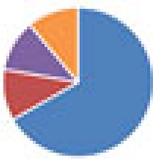
|
Mouse | Mouse(C57BL/6) |

|
3 | NR | NR | NR (119 days) | No. of IgA plasma cells per intestinal villus in duodenum, jejunum, ileum, and colon; IgA-bacterium binding in intestine; anti-E. coli IgA titer | 21 |
| ASF (8 species) | Mouse | Mouse (NMRI, C57BL/6, BALB/c, NIH Swiss, SW, NMRI, MyD88−/− Ticam1−/−, SMARTA, C57BL/6.CD45.1+) |

|
3–10 | NR | NR | NR (up to 28 days) | Cecal bacterial contents; colonic Treg cell response and relative IL-10 expression in spleen, MLN, Peyer’s patches, colonic and small intestinal LP, thoracic duct lymph; IL-17 production; relative abundance of strains; microscopic localization in colon and small intestine | 175 | |
| Mouse | Mouse (Nod1−/− and Nod2−/− on C57BL/6 background) |

|
NR | NR | NR | 6–9 wk (NR) | Cecal bacterial contents; intestinal tissue conductance and Cr-EDTA flux; E-cadherin protein expression and RegIII-gamma mRNA expression in colon; survival, colitis disease severity, histology score, and myeloperoxidase activity after DSS induction; colonic IL-6, IL-10, MCP-1, IFN-c, TNF-α, IL-12p70 levels | 17 | ||
| Mouse | Mouse (C57BL/6) |

|
NR | Autoclaved food | Both | 8–12 wk (8–12 wk) | RegIII-gamma RNA and protein expression in ileum and colon | 176 | ||
| Mouse | Mouse (C57BL/6 and C57BL/6 TSLPR−/−) |

|
3–5 | NR | NR | NR (28 days) | Expression of thymic stromal lymphopoietin mRNA in intestinal epithelial cell or colonic LP (LP); % of CD4+ T cells secreting IL-17A and IFN gamma in the colonic LP and MLN; expansion of colonic Treg cells in colonic LP and MLN; expression of receptor for TSLP by CD4+ and regulatory T cells | 177 | ||
| Mouse | Mouse (NIH Swiss) |

|
4 | NR | NR | 3 days (3 days) | Structure of myenteric plexus, nerve density, average no. of HuC/D-positive myenteric neurons per ganglion, cell body size, and average no. of nNOS-positive neurons per myenteric ganglion in duodenum, jejunum, and ileum; small intestinal motility (frequency and amplitude of muscle contractions) in duodenum, jejunum, and ileum before and after general neural or specific nitrergic blockade | 178 | ||
| Mouse | Mouse (C57BL/6) |

|
5–14 | Autoclaved mouse breeder’s diet (Harlan), unlimited access | Both | 6–12 wk (3 wk) | Colonic histology, inflammatory (MPO) activity, enteropathy (presence of fecal albumin), and cytokine expression; fecal microbiota profiles; colonic gene expression; proportion of T-cell subtypes in colonic LP and other mucosal and systemic immune compartments | 81 | ||
| ASF (8 species) (Oligo-MM12 was also used, but no host parameters were assessed) | Mouse | Mouse (C57BL/6) |

|
3 (ASF), 5–23 (Oligo-MM) | NR | Both | NR | Thicknesses of total colon and colon inner mucus (ASF); mucus turnover time (ASF); alpha diversity in colon and cecum (Oligo-MM) | 26 | |
| ASF (8 and 9 species) 8 species: ASF 9 species: ASF + Oxalobacter formigenes |
9 species: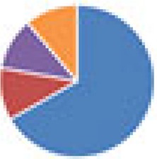
|
Mouse | Mouse (SW) |

|
4–7 | LM-485 autoclavable rodent diet, free access | M (no gender effect observed) | 3–9 mo (3–9 mo + 6 wk) | Bacterial levels in stomach, cecum, proximal colon, and cecal mucosa; body wt; dietary oxalate intake; cecal and fecal oxalate levels; urine vol; urinary metabolite levels; cecal wet wt; cecal water metabolites | 20 |
| Partial ASF (6 species) ASF356, -361, -492, -502, -519, and -500 (ASF360 and -457 did not colonize) |
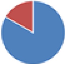
|
Mouse | Mouse (NOD.MyD88KO) | None | 9–23 | NR | Both | NR (up to 30 wk) | Incidence of diabetes; histological scores of pancreatic islet destruction | 18 |
| Partial ASF (4 and 5 species) 4 species: ASF360, ASF361, ASF457, ASF519 5 species: 4 species + Butyrivibrio fibrisolvens (type I, ATCC 19171; type II, ATCC 51255) |
4 species: 5 species: 
|
Mouse and bovine | Mouse (BALB/c) |

|
4–5 | Autoclaved low-fiber diet (5SRZ, catalog no. 1813680), high-fiber diet (5SVL, catalog no. 1813901), or tributyrin diet (5AVC, catalog no. 1814961) | NR | NR (2.5–5 mo after colorectal cancer induction) | Colorectal tumor multiplicity, tumor size, and tumor grade; levels of LDHA, lactate, butyrate, H3ac, and total H3 in colonic tissue and tumors; luminal SCFA levels; H3ac and expression levels of Fas, p21, and p27 genes in colonic tissue and tumors; apoptosis and cell proliferation levels in colonic tissue and tumors | 19 |
| Partial ASF (4, 5, 7, and 7 species) 4 species: ASF356, ASF360, ASF361, and ASF519 5 species: ASF360, ASF361, ASF457, SB2 (ASF502), and ASF519 7 species: ASF356, ASF360, ASF361, ASF457, ASF500, SB2 (ASF502), and ASF519 7 species: 4 species + E. coli Mt1B1, Streptococcus danieliae ERD01G, Staphylococcus xylosus 33-ERD13C (more with Oligo-MM [see below]) |
4 species: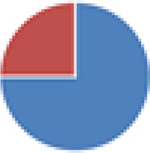 5 species: 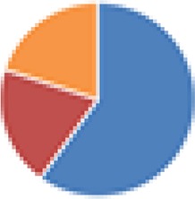 7 species: 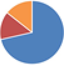 7 species: 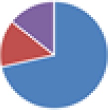
|
Mouse | Mouse (C57BL/6) |

|
4–6 | NR | Both | 0 or 8–12 wk (8–12 wk or 40 days) | Fecal bacterial content; bacterial load of S. Typhimurium in feces, cecum, and MLN; relative cecal wt; functional genomic analysis of bacteria | 22 |
| Oligo-MM (12, 15, and 17 species) 12 species, Oligo-MM: Acutalibacter muris KB18, Flavonifractor plautii YL31, Clostridium clostridioforme YL32, Blautia coccoides YL58, Clostridium innocuum I46, Lactobacillus reuteri I49, Enterococcus faecalis KB1, Bacteroides caecimuris I48, Muribaculum intestinale YL27, Bifidobacterium longum subsp. animalis YL2, Turicimonas muris YL45, Akkermansia muciniphila YL44 15 species: 12 species + 3 facultative anaerobes (E. coli Mt1B1, Streptococcus danieliae ERD01G, Staphylococcus xylosus 33-ERD13C) 17 species: 12 species + 5 ASF members (ASF360, ASF361, ASF457, SB2 [ASF502], ASF519) |
12 species: 15 species: 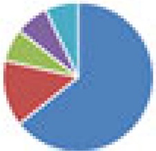 17 species: 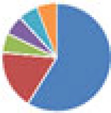
|
Mouse | Mouse (C57BL/6) |

|
4–6 | NR | Both | 0 (8–12 wk) | Fecal bacterial content; bacterial load of S. Typhimurium in feces, cecum, and MLN; relative cecal wt; functional genomic analysis of bacteria | 22 |
| Oligo-MM (12 and 13 species) 12 species: Oligo-MM 13 species: 12 species + Clostridium scindens ATCC 35704 |
12 species: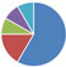 13 species: 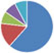
|
Mouse | Mouse (C57BL/6) |

|
5–8 | NR | NR | 0 (6–12 wk) | Fecal and cecal bacterial contents; cecal levels of lipocalin-2; calprotectin expression in cecal tissue; histopathology of cecum; cecal bile acid metabolome | |
| SIHUMI(x) (7 and 8 species): SIHUMI: Anaerostipes caccae DSM 14662 or DSM 14667, Bacteroides thetaiotaomicron DSM 2079, B. longum NCC 2705, Blautia producta DSM 2950, Clostridium ramosum DSM 1402, E. coli K-12 MG1655, Lactobacillus plantarum DSM 20174 SIHUMI(x) Clostridium butyricum DSM 10702 |
7 species: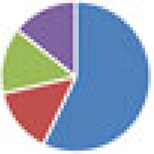 8 species: 
|
Human | Rat (Sprague-Dawley) |

|
3–21 | Sterilized standard chow (225 g/kg of body wt protein, 50 g/kg crude fat, 65 g/kg ash, 135 g/kg moisture, 480 g/kg N-free extract), fermentable-fiber-free diet, inulin diet, pectin diet, and high-fat and low-fat diets | Both | 0–3 mo (2–38 wk) | Stability of microbiota in offspring; SCFA concn and pH in cecum, colon, and feces; bacterial counts in cecum, colon, and feces; Midtvedt criteria | 27 |
| SIHUMI(x) (8 and 9 species) 8 species: SIHUMI(x) 9 species: 8 species + A. muciniphila ATCC BAA-835 |
9 species:
|
Human | Mouse (C3H) |

|
5–10 | NR | NR | 12 wk (5–15 days) | Bacterial cell no. and proportions in cecum and colon; cecal and colonic histopathology scores; expression of proinflammatory cytokines in cecal and colonic mucosa; serum protein levels of proinflammatory cytokines; no. of S. Typhimurium cells in MLN and spleen; size; macrophage infiltration in cecal tissue; localization of A. muciniphila and S. Typhimurium; mucin formation, mucus thickness, mucus composition, and no. of mucin-filled cells | 28 |
| SIHUMI(x) (8 and 9 species) 8 species: SIHUMI(x) 9 species: 8 species + Fusobacterium varium ATCC 8501 |
9 species:
|
Human | Mouse (C3H/HeOuJ) |

|
12 | Irradiated standard chow R03-40 | F | 0 (8 wk) | Body wt; dry mass of cecum and colon; bacterial content in cecum and colon; polyamine concn in cecum and colon; SCFA concn in cecum and colon; histology of cecum and distal colon (thicknesses of crypt, epithelial layer, mucosa, submucosa, muscularis externa); mitosis and apoptosis of cecal and distal colonic tissue | 30 |
| Human | Mouse (Prm/Alf, C3H/He) |

|
12–13 | Sterilized pelleted standard chow R03-40 | F | 0 (56 ± 1 days) | Lengths of small, large, and whole intestines; thicknesses of muscle, crypt, and villi in proximal and small intestine and colon; fecal and cecal microbial contents; cecal concn of SCFAs and polyamines | 31 | ||
| SIHUMI(x) (7 and 8 species) 7 species: SIHUMI(x) without C. ramosum 8 species: SIHUMI(x) |
7 species: 8 species: 
|
Human | Mouse (C3H/HeOuJ) |

|
3–9 | Irradiated low-fat or high-fat diet ad libitum | M | 0 (16 wk) | Body wt; % body fat; adipose tissue wt (epididymal, mesenteric, and subcutaneous); energy intake; food efficiency; digestibility of high-fat diet; digestible energy; cecal and colonic bacterial contents per species; blood glucose; leptin gene expression in epididymal tissue; liver wt; liver triglyceride levels; liver glycogen contents; expression of genes involved in lipid transport, lipid synthesis, cholesterol synthesis, and lipid catabolism; gene expression of proteins involved in small intestinal glucose uptake; SCFA formation in cecum, colon, and portal vein plasma; gene expression of SCFA-related proteins in colonic mucosa; gene expression of lipid transport and storage proteins in ileum; parameters of intestinal permeability and low-grade inflammation | 32 |
| SIHUMI(x) (8 and 9 species) 8 species: SIHUMI(x) 9 species: 8 species + A. muciniphila ATCC BAA-835 |
9 species:
|
Human | Mouse (C57BL/6.129P2-Il10tm1Cgn) |

|
5–6 | Irradiated standard chow (fortified type 1310; Altromin, Lage, Germany) ad libitum | M | 0 or 8 wk (3 wk) | Body wt; histopathology scores in submucosa, LP, surface epithelium, and lumen; colon length; relative mRNA levels of Tnfa, Ifng, and Reg3g; fecal lipocalin-2 concn; fecal and cecal bacterial levels; cecal histology; no. of goblet cells per 100 epithelial cells in cecum and colon; mucus layer thickness in colon; relative Muc2 mRNA levels in distal small intestine, cecum, and colon | 29 |
Abbreviations: M, male; F, female; SW, Swiss Webster; LP, lamina propria; MLN, mesenteric lymph nodes; MPO, myeloperoxidase; NR, not reported; SCFA, short-chain fatty acid; Treg cell, regulatory T cell; IL-1B, interleukin-1B; Ab, antibody; TCR, T-cell receptor; dpi, days postinfection; DSS, dextran sodium sulfate; IFN, interferon; TNF-α, tumor necrosis factor alpha; 2NT, 2-nitrotoluene; H3ac, pan-histone 3 acetylation; LDHA, lactate dehydrogenase A; nNOS, neuronal nitric oxide synthase; TgT cells, transgenic T cells; MCP-1, monocyte chemoattractant protein 1.
Two different strains tested are counted as one species. Strains were not always reported. Pathogenic species, in the case of an infection model, are not included.
The colonization time includes the time from colonization (time zero in the case of transfer of microbiota to offspring) until and including the time of sacrifice or the end of experimental (e.g., dietary) manipulations, in cases where this is clearly stated in the paper. If age is given and animals are colonized at birth, the age is included in the colonization time.
Study outcomes are reported only for animals colonized with the defined community of interest.
 , Firmicutes;
 , Bacteroidetes;
 , Actinobacteria;
 , Proteobacteria;
 , Verrucomicrobia;
 , other.
The color codes from left to right in the illustration are as follows:
 , stomach;
 , duodenum;
 , jejunum;
 , ileum;
 , cecum;
 , colon;
 , rectum;
 , feces.
