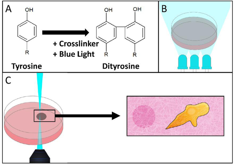
FIGURE 1.

Schematic of two crosslinking modalities: laser scanning confocal microscopy and bulk LED illumination. (A) Tyrosine residues are crosslinked to form dityrosine by blue light activated crosslinker. (B) An entire hydrogel can be crosslinked by illumination with 460 nm LEDs. (C) Illustration depicting the intended usage of selective photocrosslinking, where regions proximal to cells could be selectively stiffened by a laser scanning confocal microscope.
