
Figure 7.
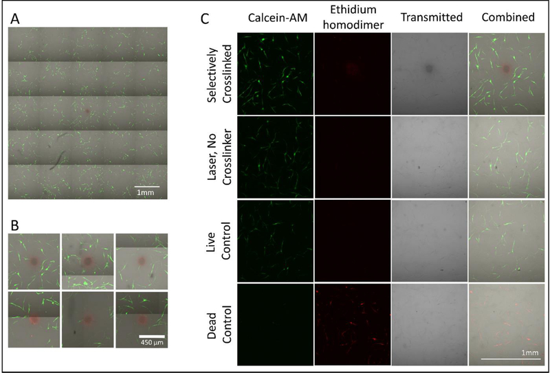
Live-dead assay of fibrin hydrogel containing NHDF cells 4 hours after T100 treatment. Calcein-AM (green) stains live cells and ethidium homodimer (red) stains dead cells. (A) Representative 5 by 5 field-of-view montage. (B) Zoomed in view around the crosslinked region for 6 replicates. (C) Areas surrounding (1) a T100 crosslinked region, (2) a control T100 without crosslinker, and (3) live and (4) dead controls. Note: The crosslinked region shows signal in the red channel even in cell-free systems with no ethidium homodimer.