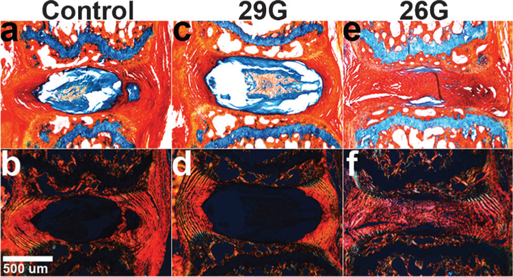
Figure 5.

Alcian Blue/Picrosirius Red stained sagittal sections of 8-week mice viewed under (a–c) brightfield and (d–f) polarized light (n = 2/group/timepoint). (a,d) Control and (b,e) 29G discs were no different at 8 weeks, while (c,f) 26G discs had collapsed height and disorganized lamellae with the presence of collagen-positive stain and lack of GAG-positive stain in the NP.
