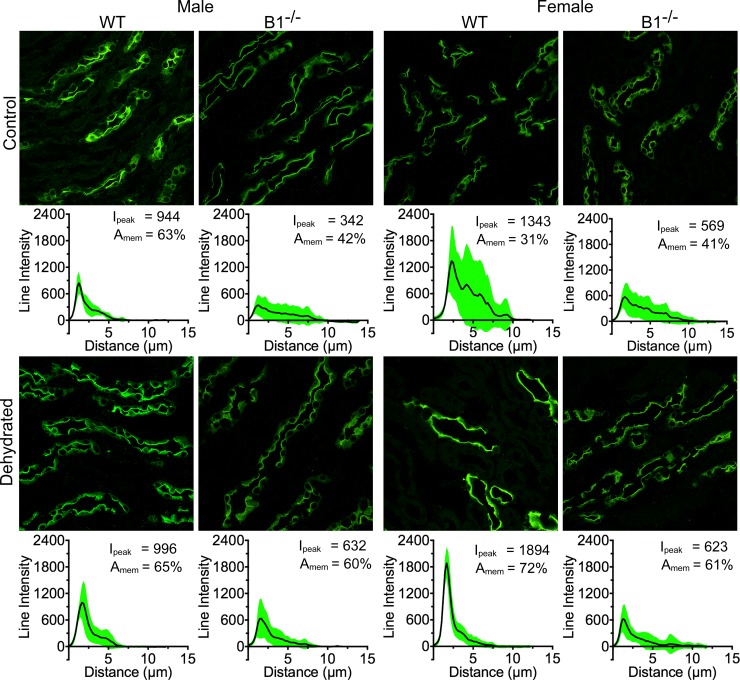
Fig 5. Female WT animals show the highest level of AQP2 regulation upon dehydration in the inner segment of the outer medulla.
WT male animals do not show much regulation in this part of the kidney (2% increase in peak intensity and AQP2 in the membrane area, 1st column), while Atp6v1b1-/- (B1-/-) males show a considerable amount of regulation with a 42% increase in AQP2 in the membrane region and an 84% increase in peak intensity (2nd column). The highest amount of regulation was seen in the case of WT female animals with a 132% increase in the AQP dependent fluorescence in the membrane region and a 41% increase in peak intensity (3rd column). Whereas female B1-/- animals showed a mere 9% increase in peak intensity, there was a 48% increase in AQP presence in the membrane region (4th column).

